• 文献检索
  • 文档翻译
  • 深度研究
  • 学术资讯
  • Suppr Zotero 插件Zotero 插件
  • 邀请有礼
  • 套餐&价格
  • 历史记录
应用&插件
Suppr Zotero 插件Zotero 插件浏览器插件Mac 客户端Windows 客户端微信小程序
定价
高级版会员购买积分包购买API积分包
服务
文献检索文档翻译深度研究API 文档MCP 服务
关于我们
关于 Suppr公司介绍联系我们用户协议隐私条款
关注我们

Suppr 超能文献

核心技术专利:CN118964589B侵权必究
粤ICP备2023148730 号-1Suppr @ 2026

文献检索

告别复杂PubMed语法,用中文像聊天一样搜索,搜遍4000万医学文献。AI智能推荐,让科研检索更轻松。

立即免费搜索

文件翻译

保留排版,准确专业,支持PDF/Word/PPT等文件格式,支持 12+语言互译。

免费翻译文档

深度研究

AI帮你快速写综述,25分钟生成高质量综述,智能提取关键信息,辅助科研写作。

立即免费体验

结核分枝杆菌中硫酸盐激活复合物激酶结构域的晶体结构。

Crystal structures of the kinase domain of the sulfate-activating complex in Mycobacterium tuberculosis.

作者信息

Poyraz Ömer, Brunner Katharina, Lohkamp Bernhard, Axelsson Hanna, Hammarström Lars G J, Schnell Robert, Schneider Gunter

机构信息

Department of Medical Biochemistry and Biophysics, Karolinska Institutet, Stockholm, Sweden.

Chemical Biology Consortium Sweden, Science for Life Laboratory Stockholm, Department of Medical Biochemistry and Biophysics, Karolinska Institutet, Stockholm, Sweden.

出版信息

PLoS One. 2015 Mar 25;10(3):e0121494. doi: 10.1371/journal.pone.0121494. eCollection 2015.

DOI:10.1371/journal.pone.0121494
PMID:25807013
原文链接:https://pmc.ncbi.nlm.nih.gov/articles/PMC4373884/
Abstract

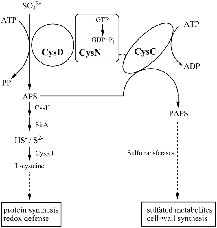
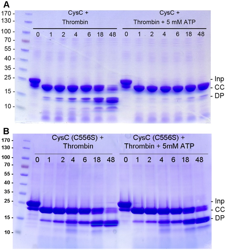
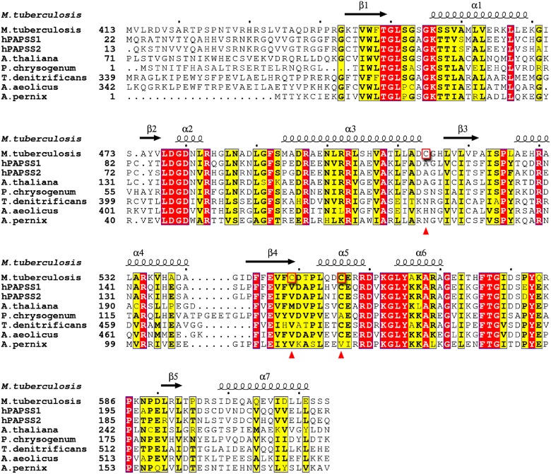
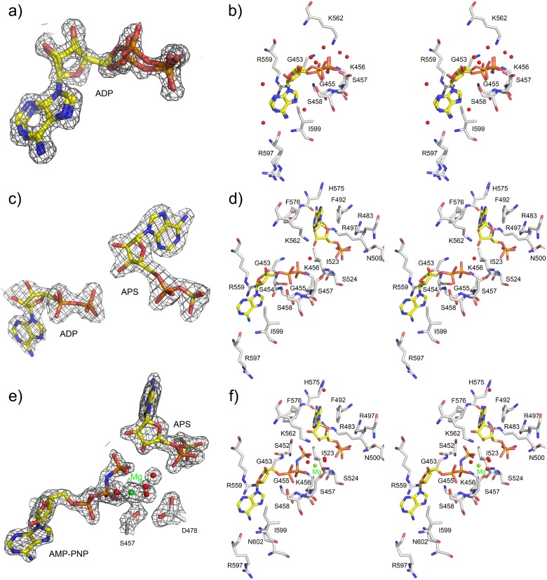
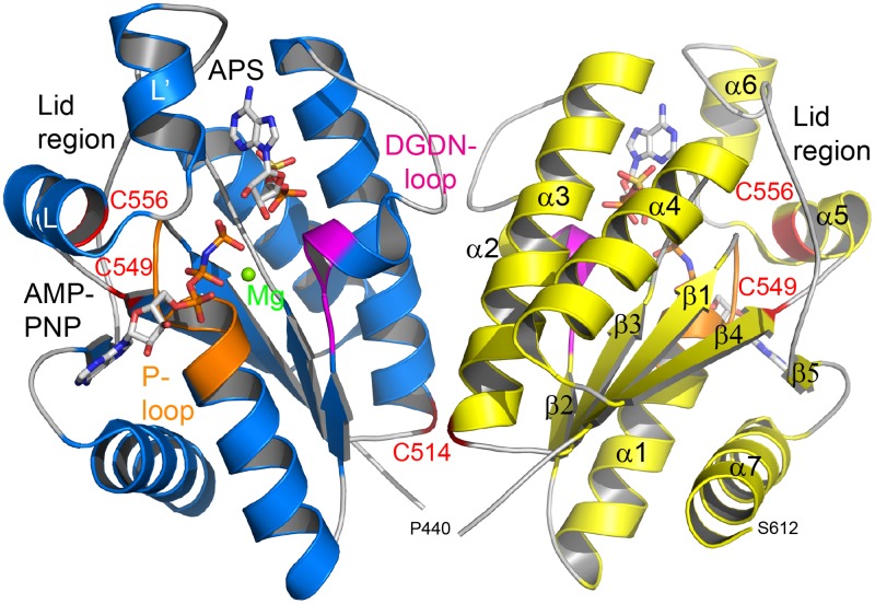
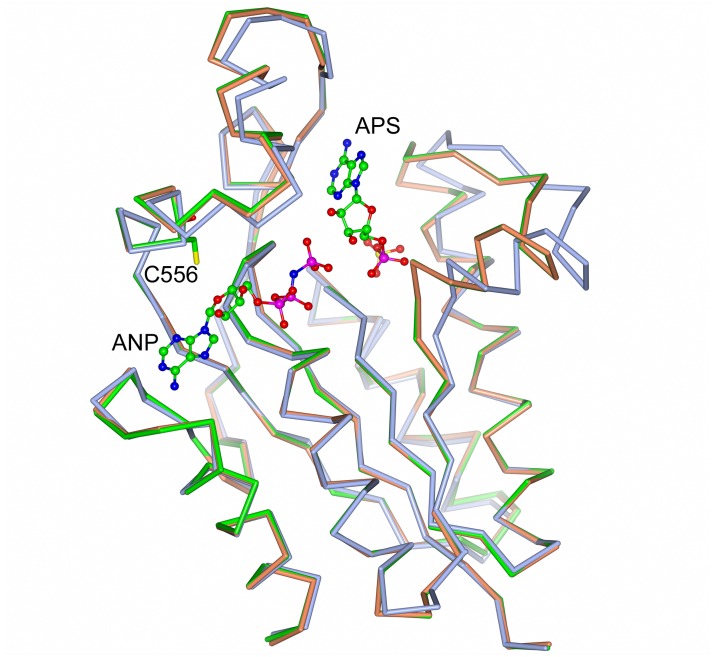
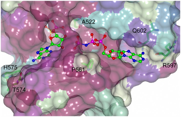
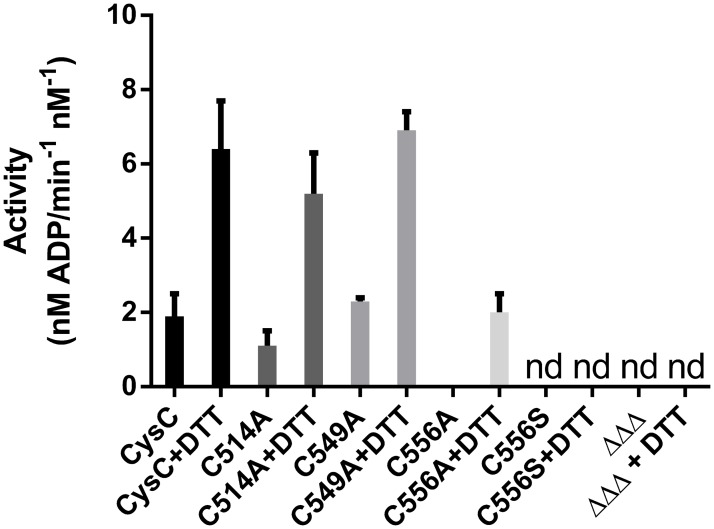
In Mycobacterium tuberculosis the sulfate activating complex provides a key branching point in sulfate assimilation. The complex consists of two polypeptide chains, CysD and CysN. CysD is an ATP sulfurylase that, with the energy provided by the GTPase activity of CysN, forms adenosine-5'-phosphosulfate (APS) which can then enter the reductive branch of sulfate assimilation leading to the biosynthesis of cysteine. The CysN polypeptide chain also contains an APS kinase domain (CysC) that phosphorylates APS leading to 3'-phosphoadenosine-5'-phosphosulfate, the sulfate donor in the synthesis of sulfolipids. We have determined the crystal structures of CysC from M. tuberculosis as a binary complex with ADP, and as ternary complexes with ADP and APS and the ATP mimic AMP-PNP and APS, respectively, to resolutions of 1.5 Å, 2.1 Å and 1.7 Å, respectively. CysC shows the typical APS kinase fold, and the structures provide comprehensive views of the catalytic machinery, conserved in this enzyme family. Comparison to the structure of the human homolog show highly conserved APS and ATP binding sites, questioning the feasibility of the design of specific inhibitors of mycobacterial CysC. Residue Cys556 is part of the flexible lid region that closes off the active site upon substrate binding. Mutational analysis revealed this residue as one of the determinants controlling lid closure and hence binding of the nucleotide substrate.

摘要

在结核分枝杆菌中,硫酸盐激活复合物是硫酸盐同化过程中的一个关键分支点。该复合物由两条多肽链CysD和CysN组成。CysD是一种ATP硫酸化酶,它利用CysN的GTPase活性提供的能量形成腺苷-5'-磷酸硫酸酯(APS),然后APS可进入硫酸盐同化的还原分支,导致半胱氨酸的生物合成。CysN多肽链还包含一个APS激酶结构域(CysC),该结构域将APS磷酸化生成3'-磷酸腺苷-5'-磷酸硫酸酯,这是硫脂合成中的硫酸盐供体。我们分别确定了结核分枝杆菌CysC与ADP形成的二元复合物、与ADP和APS形成的三元复合物以及与ATP类似物AMP-PNP和APS形成的三元复合物的晶体结构,分辨率分别为1.5 Å、2.1 Å和1.7 Å。CysC呈现出典型的APS激酶折叠结构,这些结构提供了该酶家族中保守的催化机制的全面视图。与人类同源物的结构比较显示,APS和ATP结合位点高度保守,这对设计结核分枝杆菌CysC特异性抑制剂的可行性提出了质疑。残基Cys556是柔性盖子区域的一部分,在底物结合时会封闭活性位点。突变分析表明,该残基是控制盖子关闭从而控制核苷酸底物结合的决定因素之一。

https://cdn.ncbi.nlm.nih.gov/pmc/blobs/99be/4373884/f12388d89eb0/pone.0121494.g008.jpg
https://cdn.ncbi.nlm.nih.gov/pmc/blobs/99be/4373884/6b61f0653a29/pone.0121494.g001.jpg
https://cdn.ncbi.nlm.nih.gov/pmc/blobs/99be/4373884/c7db0a783ce9/pone.0121494.g002.jpg
https://cdn.ncbi.nlm.nih.gov/pmc/blobs/99be/4373884/13d6ab5ecf53/pone.0121494.g003.jpg
https://cdn.ncbi.nlm.nih.gov/pmc/blobs/99be/4373884/0a084fd50de0/pone.0121494.g004.jpg
https://cdn.ncbi.nlm.nih.gov/pmc/blobs/99be/4373884/ce9b6447ddbb/pone.0121494.g005.jpg
https://cdn.ncbi.nlm.nih.gov/pmc/blobs/99be/4373884/850cfccc6b80/pone.0121494.g006.jpg
https://cdn.ncbi.nlm.nih.gov/pmc/blobs/99be/4373884/438f7872845d/pone.0121494.g007.jpg
https://cdn.ncbi.nlm.nih.gov/pmc/blobs/99be/4373884/f12388d89eb0/pone.0121494.g008.jpg

相似文献

1
Crystal structures of the kinase domain of the sulfate-activating complex in Mycobacterium tuberculosis.结核分枝杆菌中硫酸盐激活复合物激酶结构域的晶体结构。
PLoS One. 2015 Mar 25;10(3):e0121494. doi: 10.1371/journal.pone.0121494. eCollection 2015.
2
Crystal structure of adenosine 5'-phosphosulfate kinase isolated from Archaeoglobus fulgidus.从嗜热栖热菌中分离出的腺苷5'-磷酸硫酸激酶的晶体结构。
Biochem Biophys Res Commun. 2023 Feb 5;643:105-110. doi: 10.1016/j.bbrc.2022.12.081. Epub 2022 Dec 29.
3
5'-adenosinephosphosulfate lies at a metabolic branch point in mycobacteria.5'-腺苷磷酸硫酸酯处于分枝杆菌的一个代谢分支点上。
J Biol Chem. 2002 Sep 6;277(36):32606-15. doi: 10.1074/jbc.M204613200. Epub 2002 Jun 18.
4
Human 3'-phosphoadenosine 5'-phosphosulfate synthetase (isoform 1, brain): kinetic properties of the adenosine triphosphate sulfurylase and adenosine 5'-phosphosulfate kinase domains.人3'-磷酸腺苷5'-磷酸硫酸合成酶(同工型1,脑):三磷酸腺苷硫酸化酶和5'-磷酸腺苷硫酸激酶结构域的动力学特性
Biochemistry. 2004 Apr 13;43(14):4356-65. doi: 10.1021/bi049827m.
5
Rhizobium meliloti NodP and NodQ form a multifunctional sulfate-activating complex requiring GTP for activity.苜蓿根瘤菌NodP和NodQ形成一种多功能硫酸激活复合物,该复合物的活性需要GTP。
J Bacteriol. 1994 Nov;176(22):7055-64. doi: 10.1128/jb.176.22.7055-7064.1994.
6
Adenosine-5'-phosphosulfate--a multifaceted modulator of bifunctional 3'-phospho-adenosine-5'-phosphosulfate synthases and related enzymes.腺苷-5'-磷酸硫酸盐——双功能 3'-磷酸腺苷-5'-磷酸硫酸盐合酶及相关酶的多功能调节剂。
FEBS J. 2013 Jul;280(13):3050-7. doi: 10.1111/febs.12252. Epub 2013 Apr 17.
7
Ligand-induced structural changes in adenosine 5'-phosphosulfate kinase from Penicillium chrysogenum.来自产黄青霉的腺苷5'-磷酸硫酸激酶中配体诱导的结构变化。
Biochemistry. 2002 Nov 19;41(46):13672-80. doi: 10.1021/bi026556b.
8
Intermediate channeling between ATP sulfurylase and adenosine 5'-phosphosulfate kinase from rat chondrosarcoma.大鼠软骨肉瘤中ATP硫酸化酶与5'-磷酸腺苷硫酸激酶之间的中间通道作用
Biochemistry. 1994 Jun 7;33(22):6822-7. doi: 10.1021/bi00188a010.
9
Crystal structure of ATP sulfurylase from Penicillium chrysogenum: insights into the allosteric regulation of sulfate assimilation.产黄青霉ATP硫酸化酶的晶体结构:对硫酸盐同化变构调节的见解
Biochemistry. 2001 Jun 12;40(23):6795-804. doi: 10.1021/bi010367w.
10
Crystal structure of the bifunctional ATP sulfurylase-APS kinase from the chemolithotrophic thermophile Aquifex aeolicus.嗜热自养细菌嗜泉古菌双功能ATP硫酸化酶-APS激酶的晶体结构
J Mol Biol. 2007 Jan 19;365(3):732-43. doi: 10.1016/j.jmb.2006.10.035. Epub 2006 Oct 14.

引用本文的文献

1
Mechanism of Mongolian Medicine Batri-7 on Enteritis.蒙药巴特日-7对肠炎的作用机制
J Inflamm Res. 2025 Feb 3;18:1523-1541. doi: 10.2147/JIR.S491957. eCollection 2025.
2
CysB Is a Key Regulator of the Antifungal Activity of JK-SH007.CysB 是 JK-SH007 抗真菌活性的关键调节因子。
Int J Mol Sci. 2023 Apr 29;24(9):8067. doi: 10.3390/ijms24098067.
3
Common Mechanism of Activated Catalysis in P-loop Fold Nucleoside Triphosphatases-United in Diversity.P 环折叠核苷三磷酸酶的激活催化的共同机制——多样性统一。

本文引用的文献

1
Structural basis and evolution of redox regulation in plant adenosine-5'-phosphosulfate kinase.植物腺苷-5'-磷酸硫酸激酶氧化还原调控的结构基础与进化。
Proc Natl Acad Sci U S A. 2012 Jan 3;109(1):309-14. doi: 10.1073/pnas.1115772108. Epub 2011 Dec 19.
2
The Phenix software for automated determination of macromolecular structures.用于自动确定生物大分子结构的 Phenix 软件。
Methods. 2011 Sep;55(1):94-106. doi: 10.1016/j.ymeth.2011.07.005. Epub 2011 Jul 29.
3
Presenting your structures: the CCP4mg molecular-graphics software.
Biomolecules. 2022 Sep 22;12(10):1346. doi: 10.3390/biom12101346.
4
Commonalities of Transcriptomes in Response to Defined Persisting Macrophage Stresses.对特定持续巨噬细胞应激的转录组反应的共性。
Front Immunol. 2022 Jul 1;13:909904. doi: 10.3389/fimmu.2022.909904. eCollection 2022.
5
Identifying genetic diversity of O antigens in Aeromonas hydrophila for molecular serotype detection.鉴定嗜水气单胞菌 O 抗原的遗传多样性用于分子血清型检测。
PLoS One. 2018 Sep 5;13(9):e0203445. doi: 10.1371/journal.pone.0203445. eCollection 2018.
6
Correction: Crystal Structures of the Kinase Domain of the Sulfate-Activating Complex in Mycobacterium tuberculosis.更正:结核分枝杆菌中硫酸盐激活复合物激酶结构域的晶体结构
PLoS One. 2015 Apr 20;10(4):e0127016. doi: 10.1371/journal.pone.0127016. eCollection 2015.
展示您的结构:CCP4mg分子图形软件。
Acta Crystallogr D Biol Crystallogr. 2011 Apr;67(Pt 4):386-94. doi: 10.1107/S0907444911007281. Epub 2011 Mar 18.
4
iMOSFLM: a new graphical interface for diffraction-image processing with MOSFLM.iMOSFLM:一种用于MOSFLM衍射图像处理的新图形界面。
Acta Crystallogr D Biol Crystallogr. 2011 Apr;67(Pt 4):271-81. doi: 10.1107/S0907444910048675. Epub 2011 Mar 18.
5
The challenge of new drug discovery for tuberculosis.结核病新药研发面临的挑战。
Nature. 2011 Jan 27;469(7331):483-90. doi: 10.1038/nature09657.
6
Understanding latent tuberculosis: a moving target.理解潜伏性结核:一个动态的目标。
J Immunol. 2010 Jul 1;185(1):15-22. doi: 10.4049/jimmunol.0903856.
7
ConSurf 2010: calculating evolutionary conservation in sequence and structure of proteins and nucleic acids.ConSurf 2010:计算蛋白质和核酸序列及结构的进化保守性。
Nucleic Acids Res. 2010 Jul;38(Web Server issue):W529-33. doi: 10.1093/nar/gkq399. Epub 2010 May 16.
8
Features and development of Coot.Coot的特点与发展
Acta Crystallogr D Biol Crystallogr. 2010 Apr;66(Pt 4):486-501. doi: 10.1107/S0907444910007493. Epub 2010 Mar 24.
9
MolProbity: all-atom structure validation for macromolecular crystallography.MolProbity:用于大分子晶体学的全原子结构验证
Acta Crystallogr D Biol Crystallogr. 2010 Jan;66(Pt 1):12-21. doi: 10.1107/S0907444909042073. Epub 2009 Dec 21.
10
Structure of the two-domain hexameric APS kinase from Thiobacillus denitrificans: structural basis for the absence of ATP sulfurylase activity.反硝化硫杆菌双结构域六聚体APS激酶的结构:缺乏ATP硫酸化酶活性的结构基础。
Acta Crystallogr D Biol Crystallogr. 2009 Oct;65(Pt 10):1021-31. doi: 10.1107/S0907444909026547. Epub 2009 Sep 16.