Huang Yong, Tong Dedi, Zhu Shan, Wu Lehao, Mao Qi, Ibrahim Zuhaib, Lee W P Andrew, Brandacher Gerald, Kang Jin U
Baltimore, Md.; and Beijing, People's Republic of China From the Departments of Electrical and Computer Engineering and Plastic and Reconstructive Surgery, Johns Hopkins University; the Department of Hand Surgery, Beijing Jishuitan Hospital; and the Department of Plastic and Reconstructive Surgery, Peking Union Medical College and Chinese Academy of Medical Science.
Plast Reconstr Surg. 2015 Apr;135(4):711e-720e. doi: 10.1097/PRS.0000000000001124.
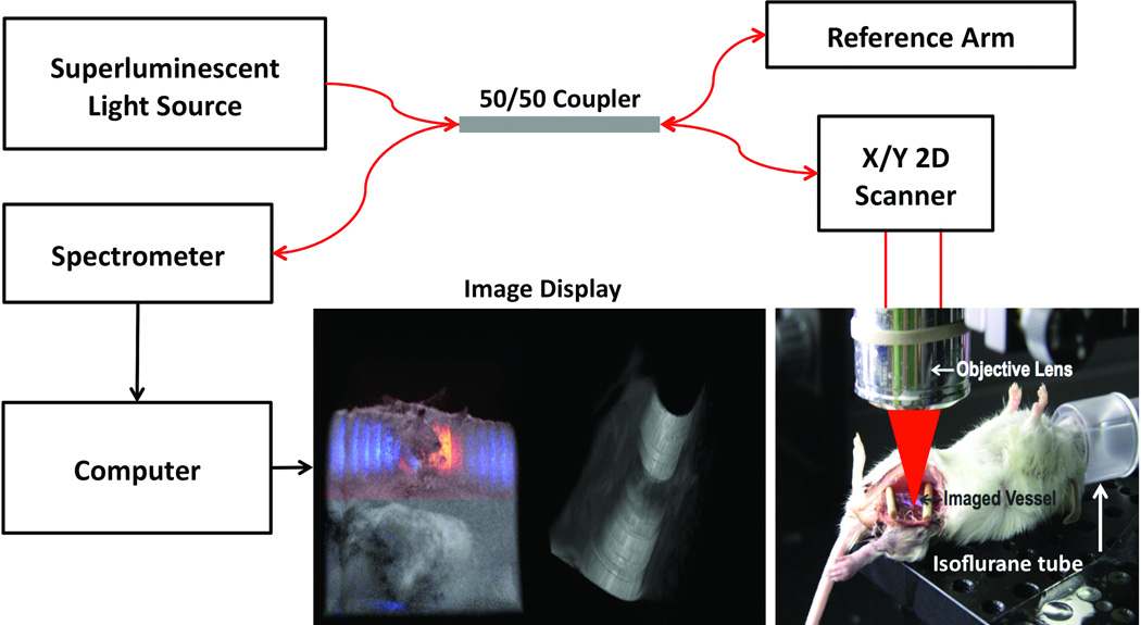
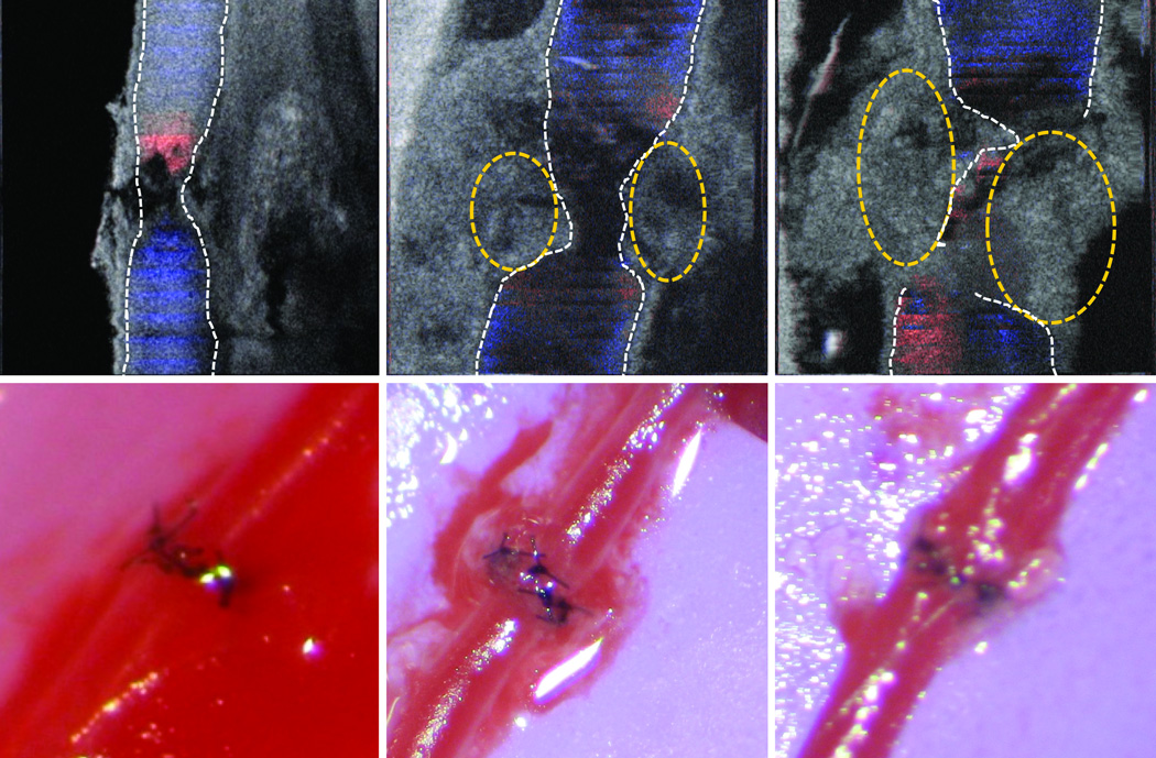
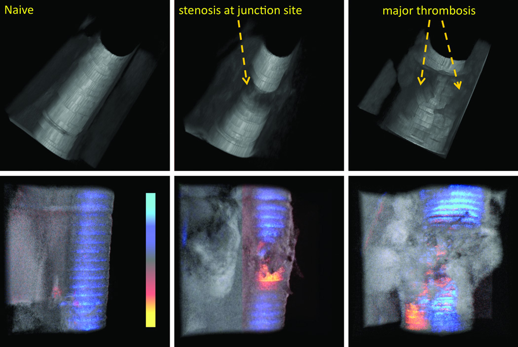
Evolution in microsurgical techniques and tools has paved the way for supermicrosurgical anastomoses, with vessel diameters often approaching below 0.8 mm in the clinical realm and even smaller (0.2 to 0.3 mm) in murine models. Several imaging and monitoring devices have been introduced for postoperative monitoring, but intraoperative guidance, assessment, and predictability have remained limited to binocular optical microscopy and the surgeon's experience. The authors present a high-resolution, real-time, three-dimensional imaging modality for intraoperative evaluation of luminal narrowing, thrombus formation, and flow alterations.
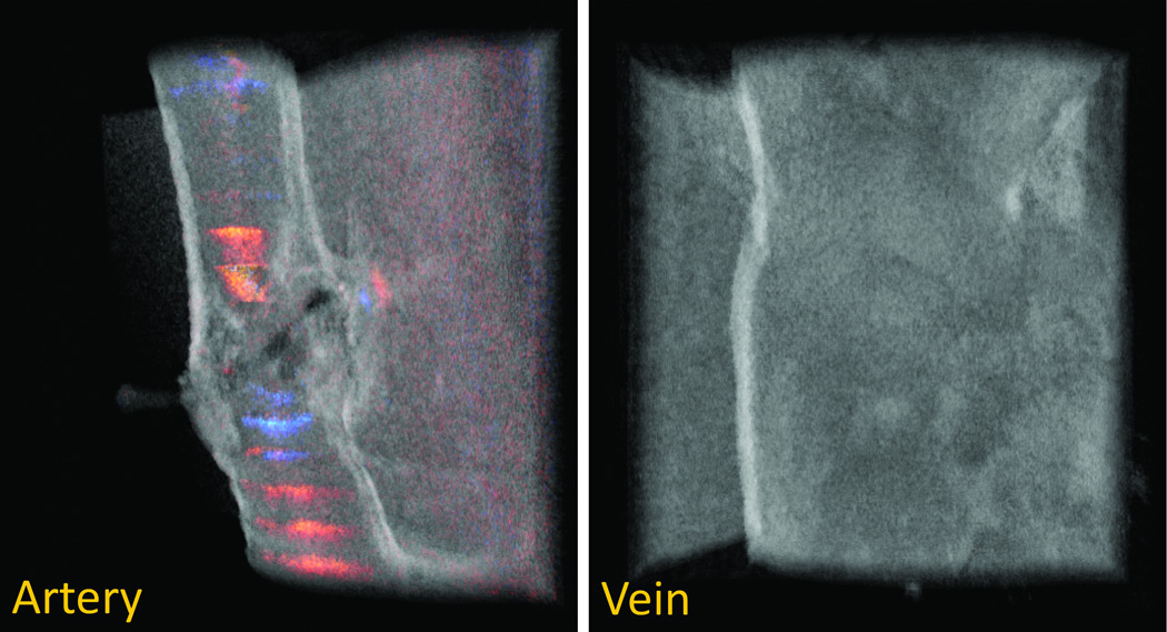
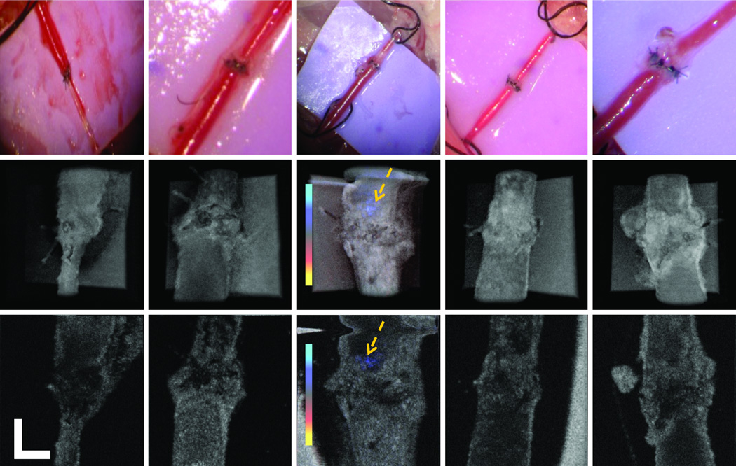
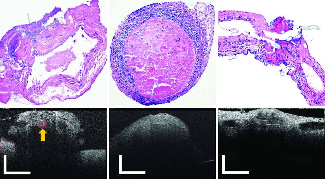
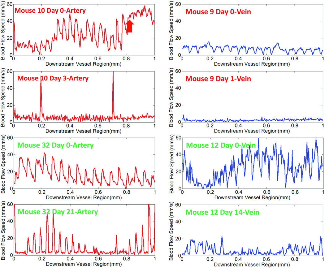
An imaging modality that provides immediate, in-depth, high-resolution, three-dimensional structure view and flow information of the anastomosed site, called phase-resolved Doppler optical coherence tomography, was developed. Twenty-two mouse femoral artery anastomoses and 17 mouse venous anastomoses were performed and evaluated. Flow status, vessel inner lumen three-dimensional structure, and early thrombus detection were analyzed based on imaging results. Predictions formed correlated with actual long-term surgical outcomes. Eventually, four cases of mouse orthotopic limb transplantation were carried out, and predicted long-term patency based on imaging results was confirmed by actual results.
The assessments based on high-resolution three-dimensional visualization of the vessel flow status and inner lumen provided by phase-resolved Doppler optical coherence tomography show 92 percent sensitivity and 90 percent specificity for arterial anastomoses and 90 percent sensitivity and 86 percent specificity for venous anastomoses.
Phase-resolved Doppler optical coherence tomography is an effective evaluation tool for microvascular anastomosis. It can predict the long-term vessel patency with high sensitivity and specificity.
显微外科技术和工具的发展为超显微外科吻合术铺平了道路,在临床领域血管直径常常接近0.8毫米以下,在鼠类模型中甚至更小(0.2至0.3毫米)。已经引入了几种成像和监测设备用于术后监测,但术中的指导、评估和可预测性仍然局限于双目光学显微镜和外科医生的经验。作者提出了一种高分辨率、实时的三维成像方式,用于术中评估管腔狭窄、血栓形成和血流改变。
开发了一种成像方式,称为相分辨多普勒光学相干断层扫描,它能提供吻合部位即时、深度、高分辨率的三维结构视图和血流信息。进行并评估了22例小鼠股动脉吻合和17例小鼠静脉吻合。根据成像结果分析血流状态、血管内腔三维结构和早期血栓检测情况。形成的预测与实际长期手术结果相关。最终,进行了4例小鼠原位肢体移植,基于成像结果预测的长期通畅情况得到了实际结果的证实。
相分辨多普勒光学相干断层扫描提供的基于血管血流状态和内腔高分辨率三维可视化的评估显示,动脉吻合的敏感性为92%,特异性为90%;静脉吻合的敏感性为90%,特异性为86%。
相分辨多普勒光学相干断层扫描是微血管吻合的一种有效评估工具。它能够以高敏感性和特异性预测血管的长期通畅情况。