Fernández-Millán Pablo, Lázaro Melisa, Cansız-Arda Şirin, Gerhold Joachim M, Rajala Nina, Schmitz Claus-A, Silva-Espiña Cristina, Gil David, Bernadó Pau, Valle Mikel, Spelbrink Johannes N, Solà Maria
Structural MitoLab; Department of Structural Biology, Molecular Biology Institute Barcelona (IBMB-CSIC), Barcelona, E-08028, Spain.
Structural Biology Unit. Centre for Cooperative Research in Biosciences, CICbioGUNE, Derio, E-48160, Spain.
Nucleic Acids Res. 2015 Apr 30;43(8):4284-95. doi: 10.1093/nar/gkv189. Epub 2015 Mar 30.
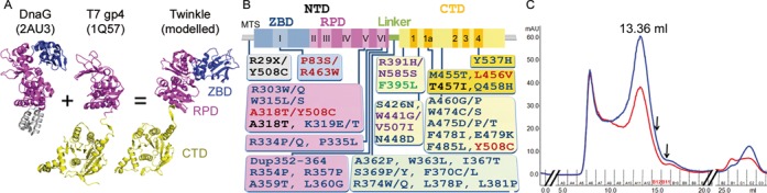
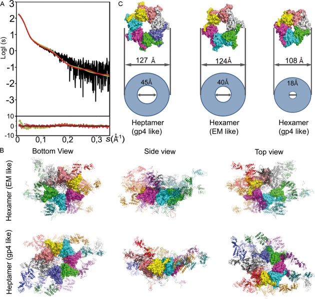
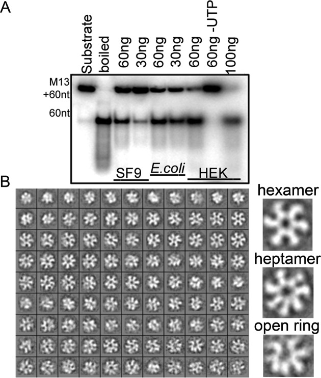
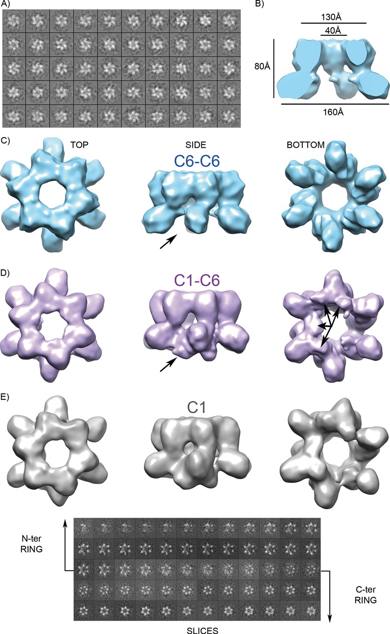
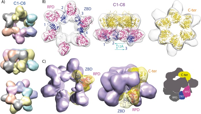
The mitochondrial replicative helicase Twinkle is involved in strand separation at the replication fork of mitochondrial DNA (mtDNA). Twinkle malfunction is associated with rare diseases that include late onset mitochondrial myopathies, neuromuscular disorders and fatal infantile mtDNA depletion syndrome. We examined its 3D structure by electron microscopy (EM) and small angle X-ray scattering (SAXS) and built the corresponding atomic models, which gave insight into the first molecular architecture of a full-length SF4 helicase that includes an N-terminal zinc-binding domain (ZBD), an intermediate RNA polymerase domain (RPD) and a RecA-like hexamerization C-terminal domain (CTD). The EM model of Twinkle reveals a hexameric two-layered ring comprising the ZBDs and RPDs in one layer and the CTDs in another. In the hexamer, contacts in trans with adjacent subunits occur between ZBDs and RPDs, and between RPDs and CTDs. The ZBDs show important structural heterogeneity. In solution, the scattering data are compatible with a mixture of extended hexa- and heptameric models in variable conformations. Overall, our structural data show a complex network of dynamic interactions that reconciles with the structural flexibility required for helicase activity.
线粒体复制解旋酶Twinkle参与线粒体DNA(mtDNA)复制叉处的链分离。Twinkle功能异常与罕见疾病相关,包括迟发性线粒体肌病、神经肌肉疾病和致命性婴儿mtDNA耗竭综合征。我们通过电子显微镜(EM)和小角X射线散射(SAXS)研究了其三维结构,并构建了相应的原子模型,这为全长SF4解旋酶的首个分子结构提供了见解,该解旋酶包括一个N端锌结合结构域(ZBD)、一个中间RNA聚合酶结构域(RPD)和一个类RecA六聚化C端结构域(CTD)。Twinkle的EM模型显示出一个六聚体双层环,一层包含ZBD和RPD,另一层包含CTD。在六聚体中,相邻亚基之间的反式接触发生在ZBD和RPD之间,以及RPD和CTD之间。ZBD表现出重要的结构异质性。在溶液中,散射数据与可变构象的延伸六聚体和七聚体模型的混合物兼容。总体而言,我们的结构数据显示了一个复杂的动态相互作用网络,这与解旋酶活性所需的结构灵活性相一致。