Ma Liangsuo, Steinberg Joel L, Keyser-Marcus Lori, Ramesh Divya, Narayana Ponnada A, Merchant Randall E, Moeller F Gerard, Cifu David X
Institute for Drug and Alcohol Studies, Virginia Commonwealth University (VCU), Richmond, VA, USA; Department of Radiology, VCU, Richmond, VA, USA.
Institute for Drug and Alcohol Studies, Virginia Commonwealth University (VCU), Richmond, VA, USA; Department of Psychiatry, VCU, Richmond, VA, USA.
Drug Alcohol Depend. 2015 Jun 1;151:128-34. doi: 10.1016/j.drugalcdep.2015.03.015. Epub 2015 Mar 23.
Diffusion tensor imaging (DTI) is a useful technique for non-invasively investigating the microstructural organization of white matter (WM), and the most consistent DTI finding regarding cocaine-related WM alterations is in the corpus callosum (CC). WM injury has also been observed in subjects with traumatic brain injury (TBI), including in the CC.
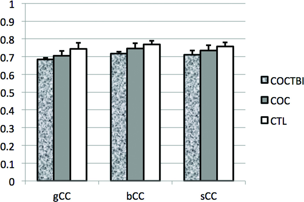
We used DTI to test if the WM microstructure is relatively more impaired in cocaine-dependent subjects who had suffered a mild TBI (mTBI). Fractional anisotropy (FA), which reflects the degree of alignment of cellular structures within fiber tracts and their structural integrity, was compared across cocaine-dependent subjects with mTBI (COCTBI group, n = 9), matched cocaine-dependent subjects without TBI (COC group, n = 12), and matched healthy controls (CTL group, n = 12).
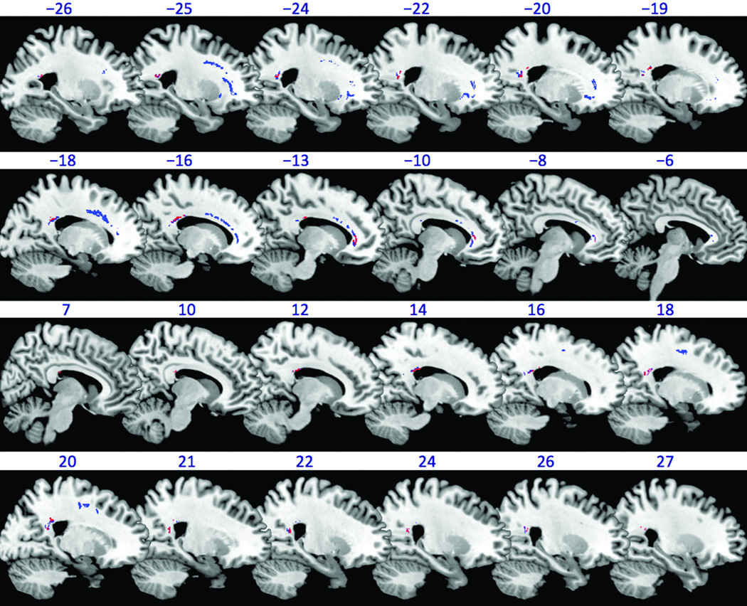
The COCTBI group had significantly lower FA in the genu, body, and splenium of CC, than the CTL group whenever the education was controlled or not. The COC group had significantly lower FA in the left and right anterior corona radiata than the CTL group only when the education was controlled. There was no significant difference in FA between the COC and COCTBI groups.
Cocaine dependence (or mTBI) related WM impairments in the CC were not detectable in this small subject sample. The significant finding in the CC suggests that the concurrence of cocaine dependence and mTBI might result in more severe damage to the CC, which could even be detected in small sample size.
扩散张量成像(DTI)是一种用于无创研究白质(WM)微观结构组织的有用技术,关于可卡因相关WM改变最一致的DTI发现是在胼胝体(CC)中。在创伤性脑损伤(TBI)患者中也观察到了WM损伤,包括在CC中。
我们使用DTI来测试在患有轻度TBI(mTBI)的可卡因依赖患者中WM微观结构是否相对受损更严重。比较了患有mTBI的可卡因依赖患者(COCTBI组,n = 9)、匹配的无TBI的可卡因依赖患者(COC组,n = 12)和匹配的健康对照者(CTL组,n = 12)的分数各向异性(FA),FA反映了纤维束内细胞结构的排列程度及其结构完整性。
无论是否控制受教育程度,COCTBI组CC膝部、体部和压部的FA均显著低于CTL组。仅在控制受教育程度时,COC组左右前放射冠的FA显著低于CTL组。COC组和COCTBI组之间的FA无显著差异。
在这个小样本受试者中未检测到可卡因依赖(或mTBI)相关的CC中WM损伤。CC中的显著发现表明,可卡因依赖和mTBI的并发可能导致CC更严重的损伤,甚至在小样本中也能检测到。