Yuan Xiao-Dong, Zhou Li-Fu, Wang Shu-Juan, Zhao Yan-Sheng, Wang Xiao-Jie, Zhang Li-Li, Wang Shou-Hong, Zhang Ya-Jie, Chen Li
Department of Neurology, Affiliated Kailuan General Hospital of Hebei United University, Tangshan, Hebei Province, China.
Department of MRI Room, Affiliated Kailuan General Hospital of Hebei United University, Tangshan, Hebei Province, China.
Neural Regen Res. 2015 Mar;10(3):490-7. doi: 10.4103/1673-5374.153701.
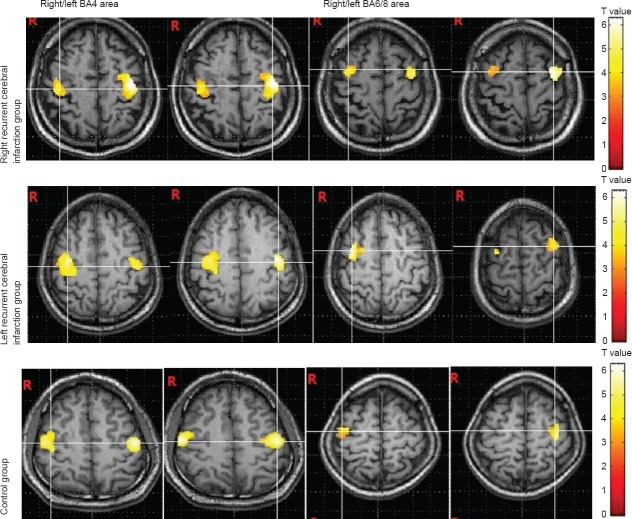
We speculate that cortical reactions evoked by swallowing activity may be abnormal in patients with central infarction with dysphagia. The present study aimed to detect functional imaging features of cerebral cortex in central dysphagia patients by using blood oxygen level-dependent functional magnetic resonance imaging techniques. The results showed that when normal controls swallowed, primary motor cortex (BA4), insula (BA13), premotor cortex (BA6/8), supramarginal gyrus (BA40), and anterior cingulate cortex (BA24/32) were activated, and that the size of the activated areas were larger in the left hemisphere compared with the right. In recurrent cerebral infarction patients with central dysphagia, BA4, BA13, BA40 and BA6/8 areas were activated, while the degree of activation in BA24/32 was decreased. Additionally, more areas were activated, including posterior cingulate cortex (BA23/31), visual association cortex (BA18/19), primary auditory cortex (BA41) and parahippocampal cortex (BA36). Somatosensory association cortex (BA7) and left cerebellum in patients with recurrent cerebral infarction with central dysphagia were also activated. Experimental findings suggest that the cerebral cortex has obvious hemisphere lateralization in response to swallowing, and patients with recurrent cerebral infarction with central dysphagia show compensatory recombination phenomena of neurological functions. In rehabilitative treatment, using the favorite food of patients can stimulate swallowing through visual, auditory, and other nerve conduction pathways, thus promoting compensatory recombination of the central cortex functions.
我们推测,吞咽活动诱发的皮质反应在伴有吞咽困难的中枢性梗死患者中可能是异常的。本研究旨在运用血氧水平依赖性功能磁共振成像技术检测中枢性吞咽困难患者大脑皮质的功能成像特征。结果显示,正常对照者吞咽时,初级运动皮质(BA4)、脑岛(BA13)、运动前皮质(BA6/8)、缘上回(BA40)和前扣带回皮质(BA24/32)被激活,且激活区域的大小左半球大于右半球。在伴有中枢性吞咽困难的复发性脑梗死患者中,BA4、BA13、BA40和BA6/8区域被激活,而BA24/32的激活程度降低。此外,更多区域被激活,包括后扣带回皮质(BA23/31)、视觉联合皮质(BA18/19)、初级听觉皮质(BA41)和海马旁皮质(BA36)。伴有中枢性吞咽困难的复发性脑梗死患者的体感联合皮质(BA7)和左侧小脑也被激活。实验结果表明,大脑皮质对吞咽反应具有明显的半球侧化,伴有中枢性吞咽困难的复发性脑梗死患者表现出神经功能的代偿重组现象。在康复治疗中,使用患者喜欢的食物可通过视觉、听觉等神经传导通路刺激吞咽,从而促进中枢皮质功能的代偿重组。