Engelhardt Sabrina, Huang Sheng-Fu, Patkar Shalmali, Gassmann Max, Ogunshola Omolara O
Fluids Barriers CNS. 2015 Feb 17;12:4. doi: 10.1186/2045-8118-12-4.
Undisturbed functioning of the blood-brain barrier (BBB) crucially depends on paracellular signaling between its associated cells; particularly endothelial cells, pericytes and astrocytes. Hypoxic and ischemic injuries are closely associated with disturbed BBB function and the contribution of perivascular cells to hypoxic/ischemic barrier regulation has gained increased attention. Regardless, detailed information on the basal hypoxic/ischemic responses of the barrier-associated cells is rare and the outcome of such cell-specific responses on BBB modulation is not well understood. This study investigated crucial parameters of hypoxic/ischemic adaptation in order to characterize individual perivascular cell responses to stress conditions.
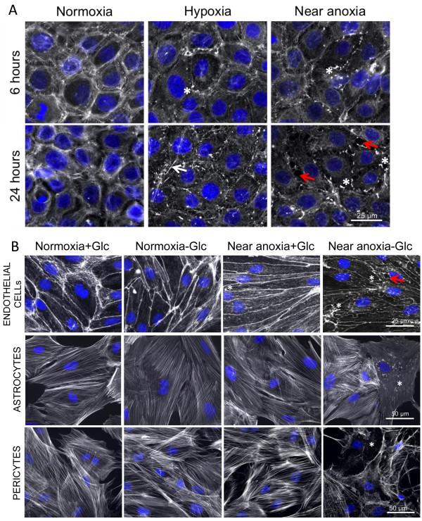
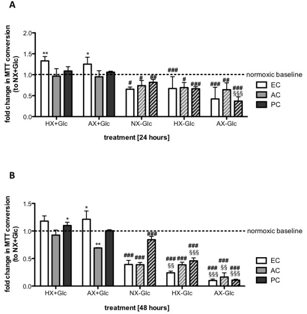
The brain microvascular endothelial cell line RBE4 (EC cell line) as well as primary rat brain endothelial cells (ECs), pericytes (PCs) and astrocytes (ACs) were exposed to 24 and 48 hours of oxygen deprivation at 1% and 0.2% O2. All primary cells were additionally subjected to combined oxygen and glucose deprivation mimicking ischemia. Central parameters of cellular adaptation and state, such as HIF-1α and HIF-1 target gene induction, actin cytoskeletal architecture, proliferation and cell viability, were compared between the cell types.
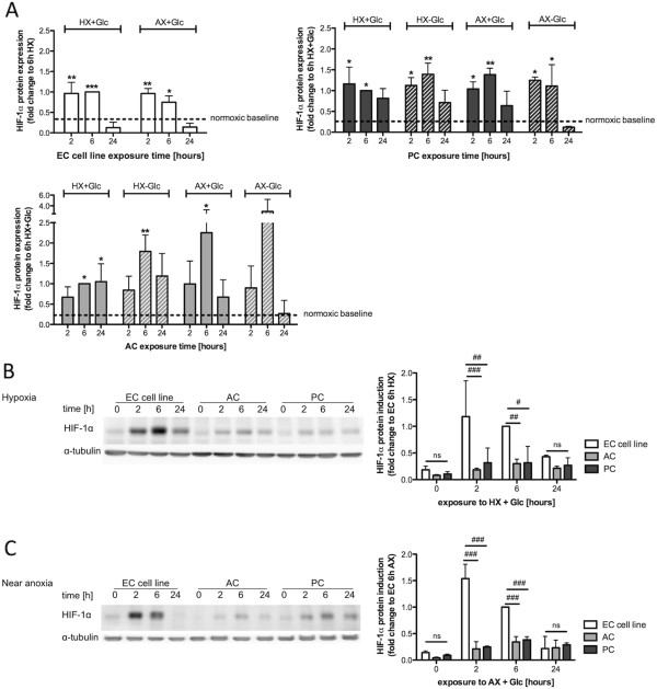
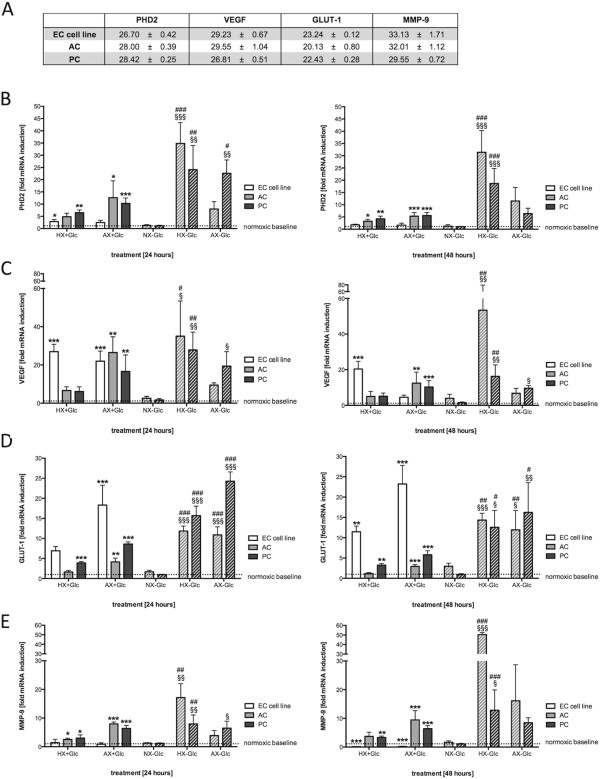
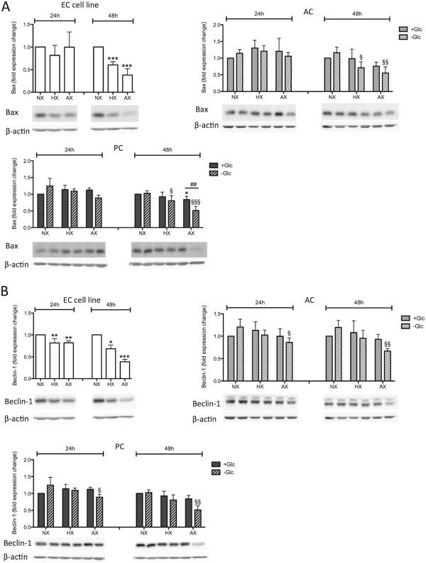
We show that endothelial cells exhibit greater responsiveness and sensitivity to oxygen deprivation than ACs and PCs. This higher sensitivity coincided with rapid and significant stabilization of HIF-1α and its downstream targets (VEGF, GLUT-1, MMP-9 and PHD2), early disruption of the actin cytoskeleton and metabolic impairment in conditions where the perivascular cells remain largely unaffected. Additional adaptation (suppression) of proliferation also likely contributes to astrocytic and pericytic tolerance during severe injury conditions. Moreover, unlike the perivascular cells, ECs were incapable of inducing autophagy (monitored via LC3-II and Beclin-1 expression) - a putative protective mechanism. Notably, both ACs and PCs were significantly more susceptible to glucose than oxygen deprivation with ACs proving to be most resistant overall.
In summary this work highlights considerable differences in sensitivity to hypoxic/ischemic injury between microvascular endothelial cells and the perivascular cells. This can have marked impact on barrier stability. Such fundamental knowledge provides an important foundation to better understand the complex cellular interactions at the BBB both physiologically and in injury-related contexts in vivo.
血脑屏障(BBB)的正常功能关键取决于其相关细胞之间的旁细胞信号传导;特别是内皮细胞、周细胞和星形胶质细胞。缺氧和缺血性损伤与BBB功能紊乱密切相关,血管周围细胞对缺氧/缺血屏障调节的作用日益受到关注。尽管如此,关于屏障相关细胞的基础缺氧/缺血反应的详细信息却很少见,而且这种细胞特异性反应对BBB调节的结果也尚未得到很好的理解。本研究调查了缺氧/缺血适应的关键参数,以表征血管周围细胞对应激条件的个体反应。
将脑微血管内皮细胞系RBE4(内皮细胞系)以及原代大鼠脑内皮细胞(ECs)、周细胞(PCs)和星形胶质细胞(ACs)分别置于1%和0.2% O₂的缺氧环境中24小时和48小时。所有原代细胞还经历了模拟缺血的联合缺氧和葡萄糖剥夺。比较了细胞类型之间细胞适应和状态的核心参数,如HIF-1α和HIF-1靶基因的诱导、肌动蛋白细胞骨架结构、增殖和细胞活力。
我们发现内皮细胞对缺氧的反应性和敏感性高于ACs和PCs。这种更高的敏感性与HIF-1α及其下游靶点(VEGF、GLUT-1、MMP-9和PHD2)的快速且显著稳定、肌动蛋白细胞骨架的早期破坏以及代谢损伤同时出现,而此时血管周围细胞基本未受影响。在严重损伤条件下,增殖的额外适应(抑制)也可能有助于星形胶质细胞和周细胞的耐受性。此外,与血管周围细胞不同,ECs无法诱导自噬(通过LC3-II和Beclin-1表达监测)——一种假定的保护机制。值得注意的是,ACs和PCs对葡萄糖剥夺的敏感性均显著高于缺氧,总体而言ACs的耐受性最强。
总之,这项工作突出了微血管内皮细胞与血管周围细胞对缺氧/缺血损伤的敏感性存在显著差异。这可能对屏障稳定性产生显著影响。这些基础知识为更好地理解体内BBB在生理和损伤相关情况下的复杂细胞相互作用提供了重要基础。