Piazzi Manuela, Williamson Andrew, Lee Chia-Fang, Pearson Stella, Lacaud Georges, Kouskoff Valerie, McCubrey James A, Cocco Lucio, Whetton Anthony D
Cell Signaling Laboratory, Department of Biomedical Science (DIBINEM), University of Bologna, Italy.
Stem Cell and Leukaemia Proteomics Laboratory, Manchester Academic Health Science Centre, The University of Manchester, Manchester, UK.
Oncotarget. 2015 May 10;6(13):10924-39. doi: 10.18632/oncotarget.3454.
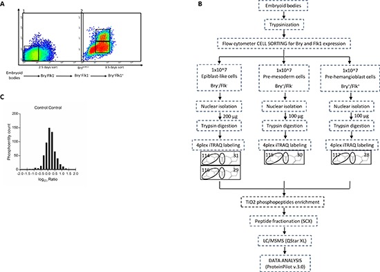
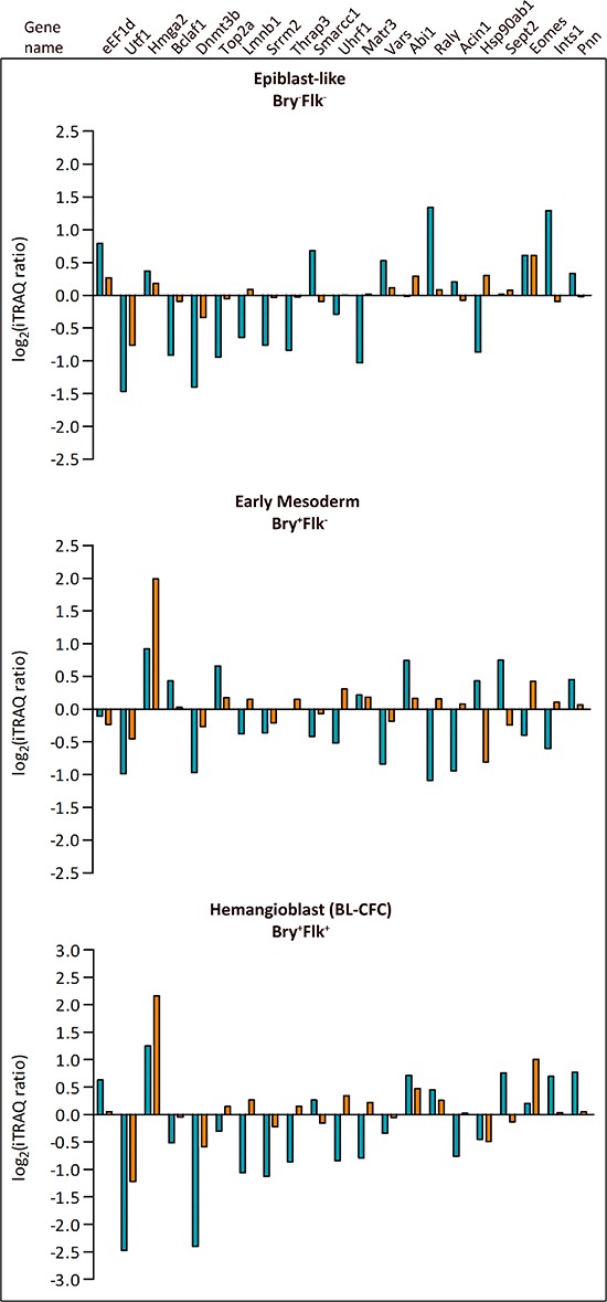
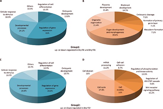
Murine embryonic stem (ES) cells can differentiate in vitro into three germ layers (endodermic, mesodermic, ectodermic). Studies on the differentiation of these cells to specific early differentiation stages has been aided by an ES cell line carrying the Green Fluorescent Protein (GFP) targeted to the Brachyury (Bry) locus which marks mesoderm commitment. Furthermore, expression of the Vascular Endothelial Growth Factor receptor 2 (Flk1) along with Bry defines hemangioblast commitment. Isobaric-tag for relative and absolute quantification (iTRAQ(TM)) and phosphopeptide enrichment coupled to liquid chromatography separation and mass spectrometry allow the study of phosphorylation changes occurring at different stages of ES cell development using Bry and Flk1 expression respectively. We identified and relatively quantified 37 phosphoentities which are modulated during mesoderm-induced ES cells differentiation, comparing epiblast-like, early mesoderm and hemangioblast-enriched cells. Among the proteins differentially phosphorylated toward mesoderm differentiation were: the epigenetic regulator Dnmt3b, the protein kinase GSK3b, the chromatin remodeling factor Smarcc1, the transcription factor Utf1; as well as protein specifically related to stem cell differentiation, as Eomes, Hmga2, Ints1 and Rif1. As most key factors regulating early hematopoietic development have also been implicated in various types of leukemia, understanding the post-translational modifications driving their regulation during normal development could result in a better comprehension of their roles during abnormal hematopoiesis in leukemia.
小鼠胚胎干细胞(ES细胞)可在体外分化为三个胚层(内胚层、中胚层、外胚层)。对这些细胞向特定早期分化阶段的分化研究,得益于一种携带靶向Brachyury(Bry)基因座的绿色荧光蛋白(GFP)的ES细胞系,该基因座标记中胚层的定向分化。此外,血管内皮生长因子受体2(Flk1)与Bry的表达共同定义了成血管细胞的定向分化。用于相对和绝对定量的等压标签(iTRAQ™)以及与液相色谱分离和质谱联用的磷酸肽富集技术,使得能够分别利用Bry和Flk1的表达,研究ES细胞发育不同阶段发生的磷酸化变化。我们鉴定并相对定量了37个磷酸化实体,这些实体在中胚层诱导的ES细胞分化过程中受到调控,比较了类上胚层细胞、早期中胚层细胞和成血管细胞富集细胞。在向中胚层分化过程中差异磷酸化的蛋白质包括:表观遗传调节因子Dnmt3b、蛋白激酶GSK3b、染色质重塑因子Smarcc1、转录因子Utf1;以及与干细胞分化特异性相关的蛋白质,如Eomes、Hmga2、Ints1和Rif1。由于大多数调节早期造血发育的关键因子也与各种类型的白血病有关,了解在正常发育过程中驱动其调控的翻译后修饰,可能有助于更好地理解它们在白血病异常造血过程中的作用。