Kalies Stefan, Antonopoulos Georgios C, Rakoski Mirko S, Heinemann Dag, Schomaker Markus, Ripken Tammo, Meyer Heiko
Biomedical Optics Department, Laser Zentrum Hannover e.V., Hanover, Germany.
Biomedical Optics Department, Laser Zentrum Hannover e.V., Hanover, Germany; Department of Cardiothoracic, Transplantation and Vascular Surgery, Hannover Medical School, Hanover, Germany.
PLoS One. 2015 Apr 24;10(4):e0124052. doi: 10.1371/journal.pone.0124052. eCollection 2015.
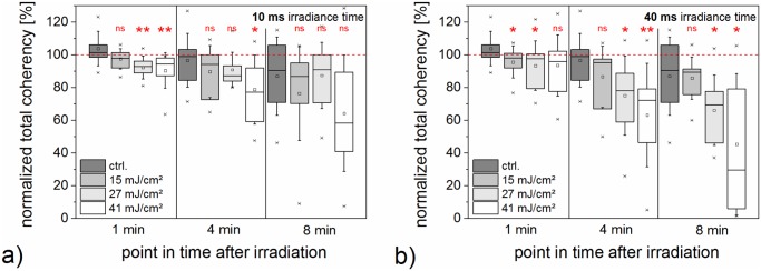
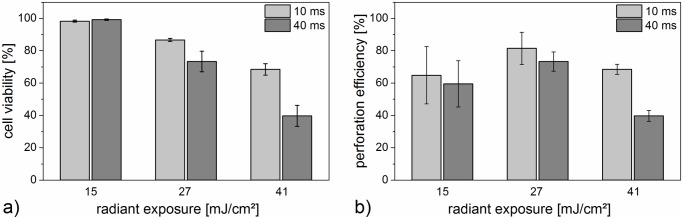
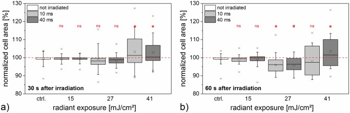
Laser based cell manipulation has proven to be a versatile tool in biomedical applications. In this context, combining weakly focused laser pulses and nanostructures, e.g. gold nanoparticles, promises to be useful for high throughput cell manipulation, such as transfection and photothermal therapy. Interactions between laser pulses and gold nanoparticles are well understood. However, it is still necessary to study cell behavior in gold nanoparticle mediated laser manipulation. While parameters like cell viability or perforation efficiency are commonly addressed, the influence of the manipulation process on other essential cell parameters is not sufficiently investigated yet. Thus, we set out to study four relevant cell properties: cell volume and area, ion exchange and cytoskeleton structure after gold nanoparticle based laser manipulation. For this, we designed a multimodal imaging and manipulation setup. 200 nm gold nanoparticles were attached unspecifically to canine cells and irradiated by weakly focused 850 ps laser pulses. Volume and area change in the first minute post laser manipulation was monitored using digital holography. Calcium imaging and cells expressing a marker for filamentous actin (F-actin) served to analyze the ion exchange and the cytoskeleton, respectively. High radiant exposures led to cells exhibiting a tendency to shrink in volume and area, possibly due to outflow of cytoplasm. An intracellular raise in calcium was observed and accompanied by an intercellular calcium wave. This multimodal approach enabled for the first time a comprehensive analysis of the cell behavior in gold nanoparticle mediated cell manipulation. Additionally, this work can pave the way for a better understanding and the evaluation of new applications in the context of cell transfection or photothermal therapy.
基于激光的细胞操作已被证明是生物医学应用中的一种多功能工具。在这种情况下,将弱聚焦激光脉冲与纳米结构(如金纳米颗粒)相结合,有望用于高通量细胞操作,如转染和光热疗法。激光脉冲与金纳米颗粒之间的相互作用已得到充分理解。然而,仍有必要研究金纳米颗粒介导的激光操作中的细胞行为。虽然细胞活力或穿孔效率等参数通常会被提及,但操作过程对其他重要细胞参数的影响尚未得到充分研究。因此,我们着手研究四个相关的细胞特性:基于金纳米颗粒的激光操作后的细胞体积和面积、离子交换和细胞骨架结构。为此,我们设计了一种多模态成像和操作装置。将200纳米的金纳米颗粒非特异性地附着在犬类细胞上,并用弱聚焦的850皮秒激光脉冲进行照射。使用数字全息术监测激光操作后第一分钟内的体积和面积变化。钙成像和表达丝状肌动蛋白(F-肌动蛋白)标记的细胞分别用于分析离子交换和细胞骨架。高辐射暴露导致细胞体积和面积有缩小的趋势,这可能是由于细胞质外流所致。观察到细胞内钙含量升高,并伴随着细胞间钙波。这种多模态方法首次实现了对金纳米颗粒介导的细胞操作中细胞行为的全面分析。此外,这项工作可为更好地理解和评估细胞转染或光热疗法背景下的新应用铺平道路。