• 文献检索
  • 文档翻译
  • 深度研究
  • 学术资讯
  • Suppr Zotero 插件Zotero 插件
  • 邀请有礼
  • 套餐&价格
  • 历史记录
应用&插件
Suppr Zotero 插件Zotero 插件浏览器插件Mac 客户端Windows 客户端微信小程序
定价
高级版会员购买积分包购买API积分包
服务
文献检索文档翻译深度研究API 文档MCP 服务
关于我们
关于 Suppr公司介绍联系我们用户协议隐私条款
关注我们

Suppr 超能文献

核心技术专利:CN118964589B侵权必究
粤ICP备2023148730 号-1Suppr @ 2026

文献检索

告别复杂PubMed语法,用中文像聊天一样搜索,搜遍4000万医学文献。AI智能推荐,让科研检索更轻松。

立即免费搜索

文件翻译

保留排版,准确专业,支持PDF/Word/PPT等文件格式,支持 12+语言互译。

免费翻译文档

深度研究

AI帮你快速写综述,25分钟生成高质量综述,智能提取关键信息,辅助科研写作。

立即免费体验

家鸭(麝香鸭)肺脏的空气传导和气体交换管道的三维结构与布局

Three-Dimensional Structure and Disposition of the Air Conducting and Gas Exchange Conduits of the Avian Lung: The Domestic Duck (Cairina moschata).

作者信息

Makanya A N, Kavoi B M, Djonov V

机构信息

Department of Veterinary Anatomy & Physiology, University of Nairobi, Riverside Drive, P.O. Box 30197, Nairobi 00100, Kenya ; Institute of Anatomy, University of Bern, Baltzerstrasse 2, 3000 Bern, Switzerland.

Department of Veterinary Anatomy & Physiology, University of Nairobi, Riverside Drive, P.O. Box 30197, Nairobi 00100, Kenya.

出版信息

ISRN Anat. 2014 Feb 5;2014:621982. doi: 10.1155/2014/621982. eCollection 2014.

DOI:10.1155/2014/621982
PMID:25938110
原文链接:https://pmc.ncbi.nlm.nih.gov/articles/PMC4392962/
Abstract

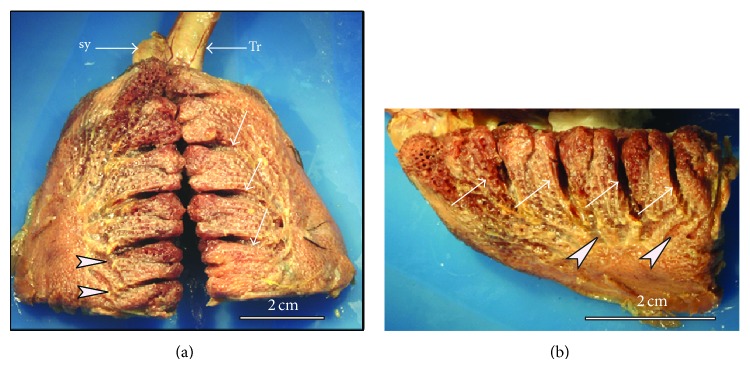
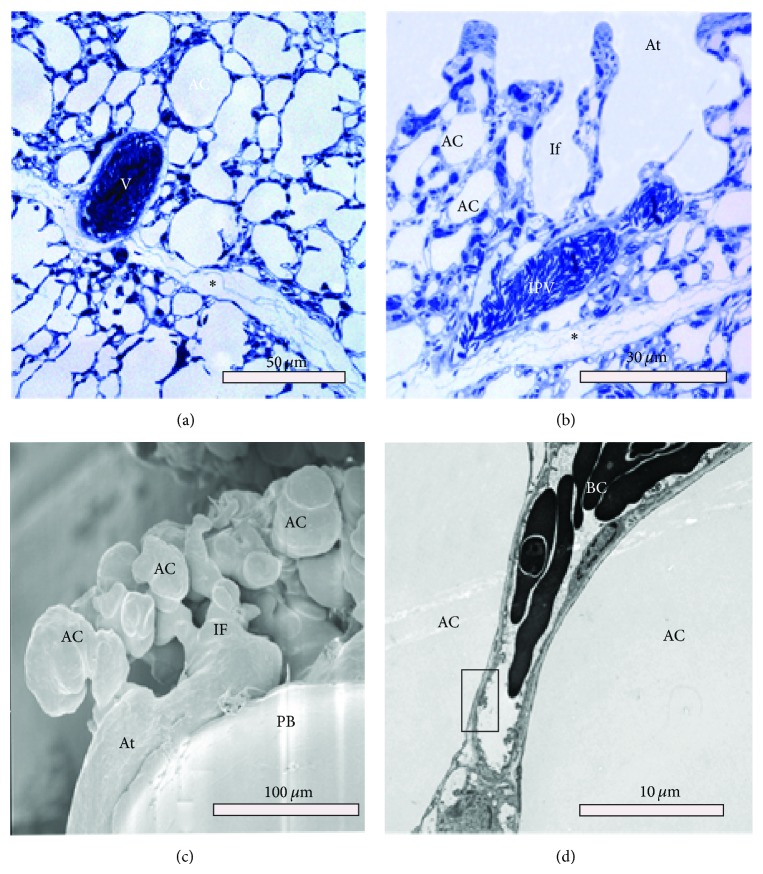
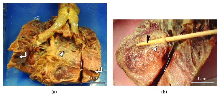
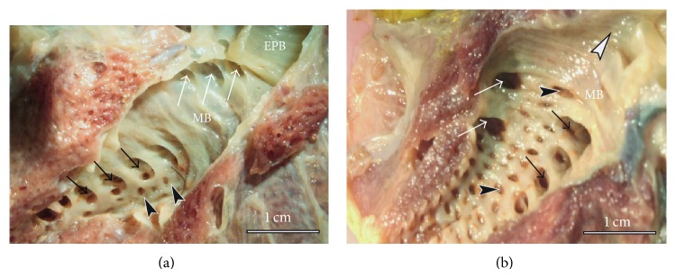
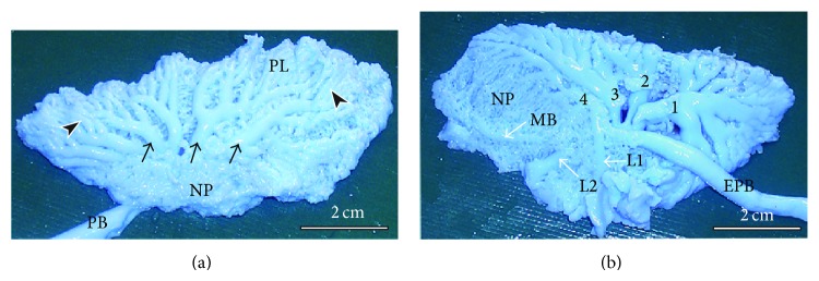
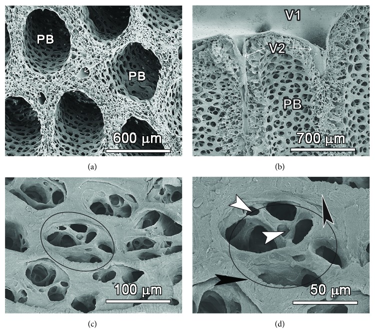
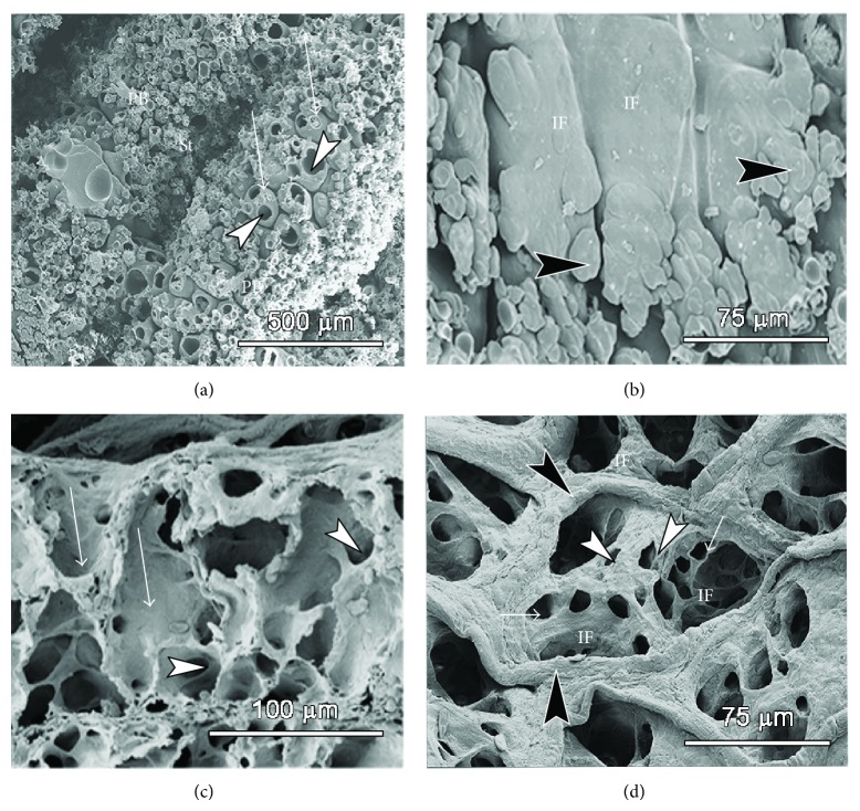
The anatomy of the domestic duck lung was studied macroscopically, by casting and by light, transmission, and scanning electron microscopy. The lung had four categories of secondary bronchi (SB), namely, the medioventral (MV, 4-5), laterodorsal (LD, 6-10), lateroventral (LV, 2-4), and posterior secondary bronchi (PO, 36-44). The neopulmonic parabronchi formed an intricate feltwork on the ventral third of the lung and inosculated those from the other SB. The lung parenchyma was organized into cylindrical parabronchi separated by thin septa containing blood vessels. Atria were shallow and well-fortified by epithelial ridges reinforced by smooth muscle bundles and gave rise to 2-6 elongate infundibulae. Air capillaries arose either directly from the atria or from infundibulae and were tubular or globular in shape with thin interconnecting branches. The newly described spatial disposition of the conducting air conduits closely resembles that of the chicken. This remarkable similarity between the categories, numbers, and 3D arrangement of the SB in the duck and chicken points to a convergence in function-oriented design. To illuminate airflow dynamics in the avian lung, precise directions of airflow in the various categories of SB and parabronchi need to be characterized.

摘要

通过铸型、光学显微镜、透射电子显微镜和扫描电子显微镜对家鸭肺的解剖结构进行了宏观研究。肺有四类二级支气管(SB),即中腹支气管(MV,4 - 5条)、背外侧支气管(LD,6 - 10条)、腹外侧支气管(LV,2 - 4条)和后二级支气管(PO,36 - 44条)。新肺段支气管在肺腹侧三分之一处形成复杂的网状结构,并与其他二级支气管的支气管相互吻合。肺实质由被含血管的薄隔膜分隔的圆柱形支气管组成。心房浅,由平滑肌束加强的上皮嵴充分加固,并产生2 - 6个细长的漏斗部。气毛细血管直接起源于心房或漏斗部,呈管状或球状,有细的相互连接的分支。新描述的传导气道的空间布局与鸡的非常相似。鸭和鸡的二级支气管在类别、数量和三维排列上的这种显著相似性表明在功能导向设计上存在趋同现象。为了阐明鸟类肺中的气流动力学,需要确定各类二级支气管和支气管中气流的精确方向。

https://cdn.ncbi.nlm.nih.gov/pmc/blobs/e9ce/4392962/9f25200095ba/ISRN.ANATOMY2014-621982.007.jpg
https://cdn.ncbi.nlm.nih.gov/pmc/blobs/e9ce/4392962/04b4ca4ef571/ISRN.ANATOMY2014-621982.001.jpg
https://cdn.ncbi.nlm.nih.gov/pmc/blobs/e9ce/4392962/7c9cd57c6ea2/ISRN.ANATOMY2014-621982.002.jpg
https://cdn.ncbi.nlm.nih.gov/pmc/blobs/e9ce/4392962/893798657225/ISRN.ANATOMY2014-621982.003.jpg
https://cdn.ncbi.nlm.nih.gov/pmc/blobs/e9ce/4392962/f3283876e577/ISRN.ANATOMY2014-621982.004.jpg
https://cdn.ncbi.nlm.nih.gov/pmc/blobs/e9ce/4392962/ea5dabfde8eb/ISRN.ANATOMY2014-621982.005.jpg
https://cdn.ncbi.nlm.nih.gov/pmc/blobs/e9ce/4392962/e07062896d8d/ISRN.ANATOMY2014-621982.006.jpg
https://cdn.ncbi.nlm.nih.gov/pmc/blobs/e9ce/4392962/9f25200095ba/ISRN.ANATOMY2014-621982.007.jpg

相似文献

1
Three-Dimensional Structure and Disposition of the Air Conducting and Gas Exchange Conduits of the Avian Lung: The Domestic Duck (Cairina moschata).家鸭(麝香鸭)肺脏的空气传导和气体交换管道的三维结构与布局
ISRN Anat. 2014 Feb 5;2014:621982. doi: 10.1155/2014/621982. eCollection 2014.
2
Development and spatial organization of the air conduits in the lung of the domestic fowl, Gallus gallus variant domesticus.家鸡(原鸡变种家鸡)肺中气道的发育与空间组织
Microsc Res Tech. 2008 Sep;71(9):689-702. doi: 10.1002/jemt.20608.
3
Anatomical and Functional Study of the Ostrich () Lung through Macroscopic Analysis in Combination with Optical and Electron Microscopy Techniques.通过宏观分析结合光学和电子显微镜技术对鸵鸟肺进行的解剖学和功能研究。
Animals (Basel). 2024 Jan 19;14(2):316. doi: 10.3390/ani14020316.
4
Spatial and functional relationships between air conduits and blood capillaries in the pulmonary gas exchange tissue of adult and developing chickens.成鸡和雏鸡肺气体交换组织中空气导管与毛细血管的空间和功能关系。
Microsc Res Tech. 2011 Feb;74(2):159-69. doi: 10.1002/jemt.20887.
5
A systematic study of the development of the airway (bronchial) system of the avian lung from days 3 to 26 of embryogenesis: a transmission electron microscopic study on the domestic fowl, Gallus gallus variant domesticus.对家鸡(原鸡变种家鸡)胚胎发育第3天至26天期间肺气道(支气管)系统发育的系统研究:透射电子显微镜研究
Tissue Cell. 2003 Oct;35(5):375-91. doi: 10.1016/s0040-8166(03)00058-2.
6
Parabronchial angioarchitecture in developing and adult chickens.发育中和成年鸡的支气管旁血管结构
J Appl Physiol (1985). 2009 Jun;106(6):1959-69. doi: 10.1152/japplphysiol.91570.2008. Epub 2009 Mar 26.
7
Slight volume changes in the duck lung do not imply a fundamental change in the structure of the parenchyma.鸭肺的轻微体积变化并不意味着实质结构发生了根本性变化。
Anat Histol Embryol. 2021 Jan;50(1):169-174. doi: 10.1111/ahe.12615. Epub 2020 Sep 24.
8
Pivotal debates and controversies on the structure and function of the avian respiratory system: setting the record straight.关于鸟类呼吸系统结构和功能的关键争论和争议:澄清事实。
Biol Rev Camb Philos Soc. 2017 Aug;92(3):1475-1504. doi: 10.1111/brv.12292. Epub 2016 Jul 28.
9
A 3D digital reconstruction of the components of the gas exchange tissue of the lung of the muscovy duck, Cairina moschata.番鸭(Cairina moschata)肺气体交换组织各组成部分的三维数字重建。
J Anat. 2005 May;206(5):477-92. doi: 10.1111/j.1469-7580.2005.00413.x.
10
Relationship of structure and function of the avian respiratory system to disease susceptibility.鸟类呼吸系统的结构与功能和疾病易感性的关系。
Poult Sci. 1998 Aug;77(8):1130-8. doi: 10.1093/ps/77.8.1130.

引用本文的文献

1
Anatomical and Functional Study of the Ostrich () Lung through Macroscopic Analysis in Combination with Optical and Electron Microscopy Techniques.通过宏观分析结合光学和电子显微镜技术对鸵鸟肺进行的解剖学和功能研究。
Animals (Basel). 2024 Jan 19;14(2):316. doi: 10.3390/ani14020316.

本文引用的文献

1
Pulmonary anatomy in the Nile crocodile and the evolution of unidirectional airflow in Archosauria.尼罗鳄的肺部解剖结构与初龙类中单向气流的演化。
PeerJ. 2013 Mar 26;1:e60. doi: 10.7717/peerj.60. Print 2013.
2
Development and remodeling of the vertebrate blood-gas barrier.脊椎动物血气屏障的发育和重塑。
Biomed Res Int. 2013;2013:101597. doi: 10.1155/2013/101597. Epub 2012 Dec 27.
3
Renal expression and function of oat1 and oat3 in rats with vascular calcification.大鼠血管钙化模型中 oat1 和 oat3 的肾表达和功能。
Pharmacology. 2012;90(1-2):66-77. doi: 10.1159/000339448. Epub 2012 Jun 29.
4
The pulmonary blood-gas barrier in the avian embryo: inauguration, development and refinement.禽类胚胎的肺气血屏障:启动、发育和完善。
Respir Physiol Neurobiol. 2011 Aug 31;178(1):30-8. doi: 10.1016/j.resp.2011.03.028. Epub 2011 Apr 6.
5
Spatial and functional relationships between air conduits and blood capillaries in the pulmonary gas exchange tissue of adult and developing chickens.成鸡和雏鸡肺气体交换组织中空气导管与毛细血管的空间和功能关系。
Microsc Res Tech. 2011 Feb;74(2):159-69. doi: 10.1002/jemt.20887.
6
Unidirectional airflow in the lungs of alligators.肺内的单向气流在鳄鱼中。
Science. 2010 Jan 15;327(5963):338-40. doi: 10.1126/science.1180219.
7
Three-dimensional serial section computer reconstruction of the arrangement of the structural components of the parabronchus of the Ostrich, Struthio camelus lung.鸵鸟肺副支气管结构成分的三维连续切片计算机重建。
Anat Rec (Hoboken). 2009 Nov;292(11):1685-98. doi: 10.1002/ar.21002.
8
Parabronchial angioarchitecture in developing and adult chickens.发育中和成年鸡的支气管旁血管结构
J Appl Physiol (1985). 2009 Jun;106(6):1959-69. doi: 10.1152/japplphysiol.91570.2008. Epub 2009 Mar 26.
9
Development and spatial organization of the air conduits in the lung of the domestic fowl, Gallus gallus variant domesticus.家鸡(原鸡变种家鸡)肺中气道的发育与空间组织
Microsc Res Tech. 2008 Sep;71(9):689-702. doi: 10.1002/jemt.20608.
10
Study of the structure of the air and blood capillaries of the gas exchange tissue of the avian lung by serial section three-dimensional reconstruction.通过连续切片三维重建对鸟类肺气体交换组织的空气毛细血管和血液毛细血管结构的研究。
J Microsc. 2008 Apr;230(Pt 1):84-93. doi: 10.1111/j.1365-2818.2008.01958.x.