• 文献检索
  • 文档翻译
  • 深度研究
  • 学术资讯
  • Suppr Zotero 插件Zotero 插件
  • 邀请有礼
  • 套餐&价格
  • 历史记录
应用&插件
Suppr Zotero 插件Zotero 插件浏览器插件Mac 客户端Windows 客户端微信小程序
定价
高级版会员购买积分包购买API积分包
服务
文献检索文档翻译深度研究API 文档MCP 服务
关于我们
关于 Suppr公司介绍联系我们用户协议隐私条款
关注我们

Suppr 超能文献

核心技术专利:CN118964589B侵权必究
粤ICP备2023148730 号-1Suppr @ 2026

文献检索

告别复杂PubMed语法,用中文像聊天一样搜索,搜遍4000万医学文献。AI智能推荐,让科研检索更轻松。

立即免费搜索

文件翻译

保留排版,准确专业,支持PDF/Word/PPT等文件格式,支持 12+语言互译。

免费翻译文档

深度研究

AI帮你快速写综述,25分钟生成高质量综述,智能提取关键信息,辅助科研写作。

立即免费体验

心外膜再生受心脏流出道和刺猬信号通路的引导。

Epicardial regeneration is guided by cardiac outflow tract and Hedgehog signalling.

作者信息

Wang Jinhu, Cao Jingli, Dickson Amy L, Poss Kenneth D

机构信息

Department of Cell Biology and Howard Hughes Medical Institute, Duke University Medical Center, Durham, NC 27710, USA.

出版信息

Nature. 2015 Jun 11;522(7555):226-230. doi: 10.1038/nature14325. Epub 2015 May 4.

DOI:10.1038/nature14325
PMID:25938716
原文链接:https://pmc.ncbi.nlm.nih.gov/articles/PMC4494087/
Abstract

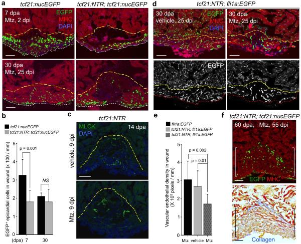
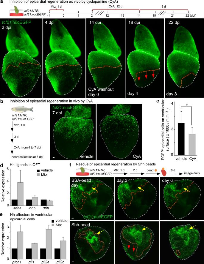
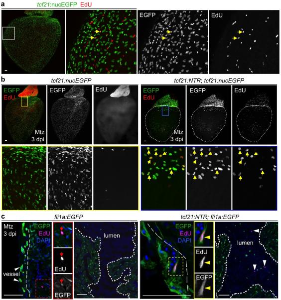
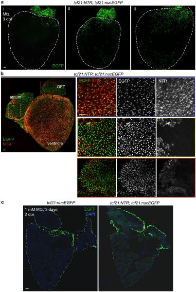
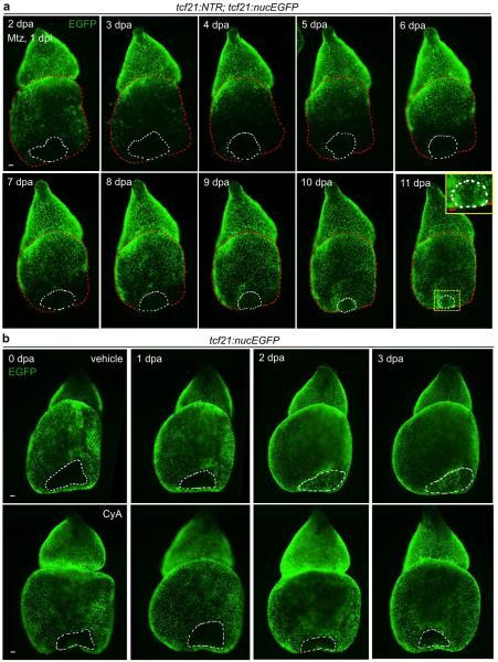
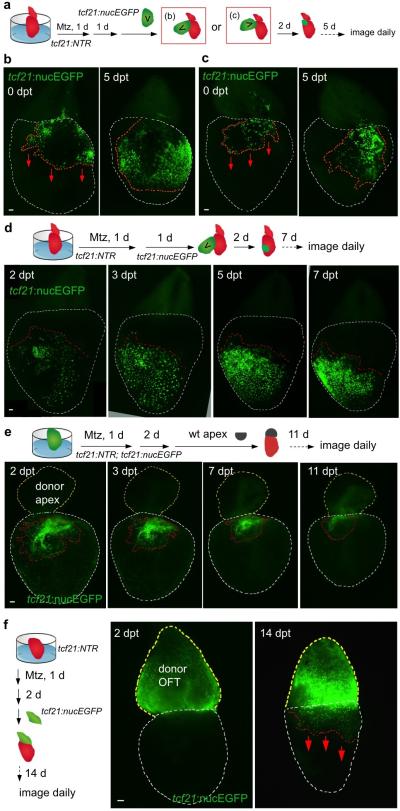
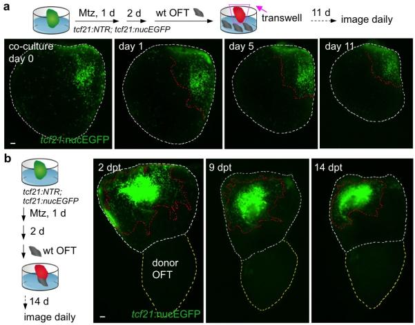
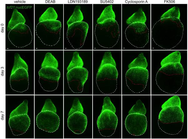
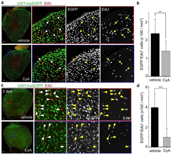
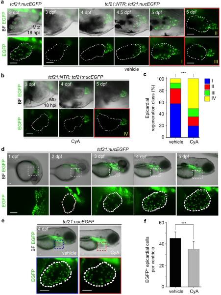
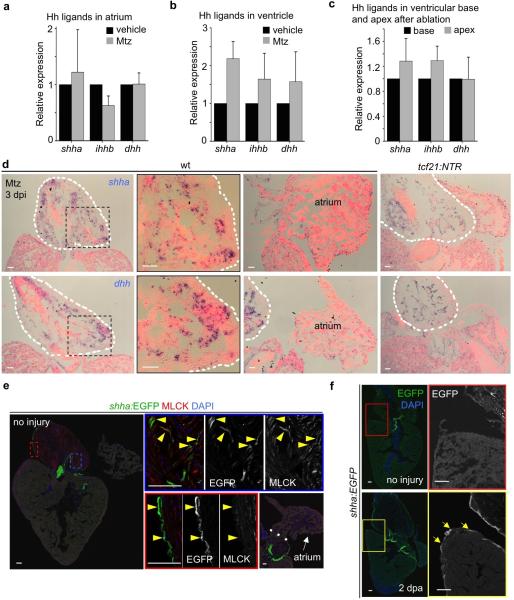
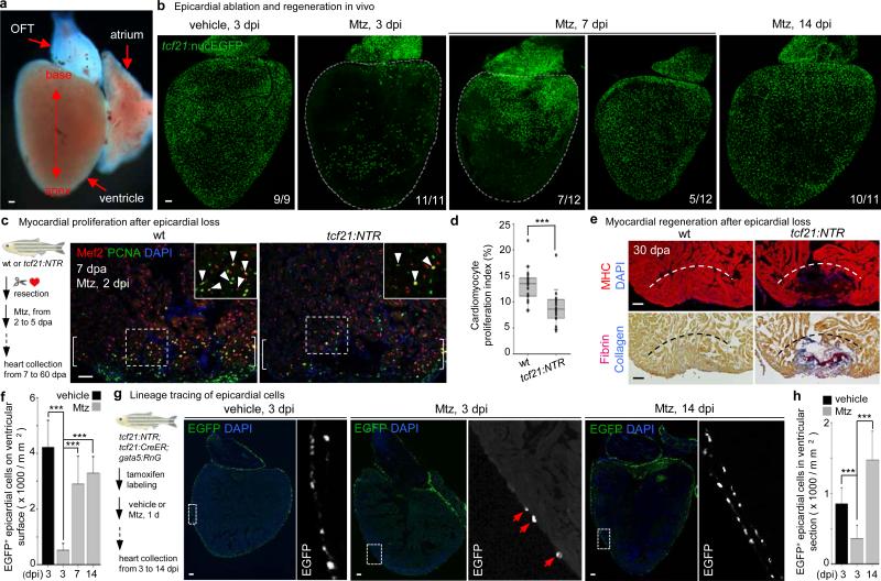
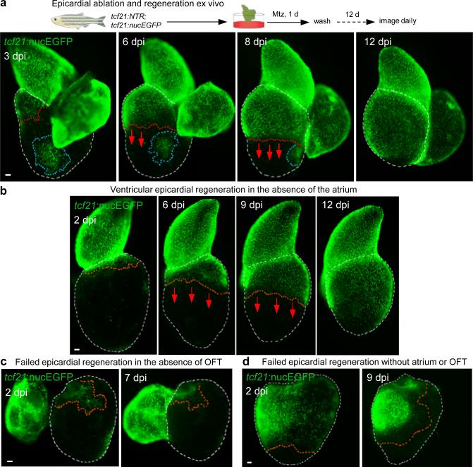
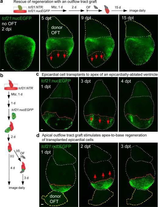
In response to cardiac damage, a mesothelial tissue layer enveloping the heart called the epicardium is activated to proliferate and accumulate at the injury site. Recent studies have implicated the epicardium in multiple aspects of cardiac repair: as a source of paracrine signals for cardiomyocyte survival or proliferation; a supply of perivascular cells and possibly other cell types such as cardiomyocytes; and as a mediator of inflammation. However, the biology and dynamism of the adult epicardium is poorly understood. To investigate this, we created a transgenic line to ablate the epicardial cell population in adult zebrafish. Here we find that genetic depletion of the epicardium after myocardial loss inhibits cardiomyocyte proliferation and delays muscle regeneration. The epicardium vigorously regenerates after its ablation, through proliferation and migration of spared epicardial cells as a sheet to cover the exposed ventricular surface in a wave from the chamber base towards its apex. By reconstituting epicardial regeneration ex vivo, we show that extirpation of the bulbous arteriosus-a distinct, smooth-muscle-rich tissue structure that distributes outflow from the ventricle-prevents epicardial regeneration. Conversely, experimental repositioning of the bulbous arteriosus by tissue recombination initiates epicardial regeneration and can govern its direction. Hedgehog (Hh) ligand is expressed in the bulbous arteriosus, and treatment with a Hh signalling antagonist arrests epicardial regeneration and blunts the epicardial response to muscle injury. Transplantation of Sonic hedgehog (Shh)-soaked beads at the ventricular base stimulates epicardial regeneration after bulbous arteriosus removal, indicating that Hh signalling can substitute for the influence of the outflow tract. Thus, the ventricular epicardium has pronounced regenerative capacity, regulated by the neighbouring cardiac outflow tract and Hh signalling. These findings extend our understanding of tissue interactions during regeneration and have implications for mobilizing epicardial cells for therapeutic heart repair.

摘要

为响应心脏损伤,包裹心脏的间皮组织层(称为心外膜)被激活,在损伤部位增殖并聚集。最近的研究表明,心外膜在心脏修复的多个方面发挥作用:作为旁分泌信号的来源,促进心肌细胞存活或增殖;提供血管周围细胞以及可能的其他细胞类型,如心肌细胞;并作为炎症的介质。然而,成人心外膜的生物学特性和动态变化仍知之甚少。为了研究这一点,我们创建了一个转基因品系,用于在成年斑马鱼中消融心外膜细胞群。在此我们发现,心肌损失后心外膜的基因缺失会抑制心肌细胞增殖并延迟肌肉再生。心外膜在消融后能有力地再生,通过留存的心外膜细胞作为一个整体进行增殖和迁移,以波浪形式从心室基部向心尖覆盖暴露的心室表面。通过在体外重建心外膜再生,我们发现摘除球囊动脉(一种独特的、富含平滑肌的组织结构,负责分配心室流出的血液)会阻止心外膜再生。相反,通过组织重组对球囊动脉进行实验性重新定位可启动心外膜再生并控制其方向。刺猬(Hh)配体在球囊动脉中表达,用Hh信号拮抗剂处理会阻止心外膜再生,并减弱心外膜对肌肉损伤的反应。在心室基部移植浸泡过音猬因子(Shh)的珠子可刺激球囊动脉摘除后的心外膜再生,表明Hh信号可替代流出道的影响。因此,心室心外膜具有显著的再生能力,受邻近的心脏流出道和Hh信号调控。这些发现扩展了我们对再生过程中组织相互作用的理解,并对动员心外膜细胞进行治疗性心脏修复具有启示意义。

https://cdn.ncbi.nlm.nih.gov/pmc/blobs/f078/4494087/7460d9d1dd08/nihms-664241-f0004.jpg
https://cdn.ncbi.nlm.nih.gov/pmc/blobs/f078/4494087/9c4d47be721f/nihms-664241-f0005.jpg
https://cdn.ncbi.nlm.nih.gov/pmc/blobs/f078/4494087/995c9116dbb0/nihms-664241-f0006.jpg
https://cdn.ncbi.nlm.nih.gov/pmc/blobs/f078/4494087/d065a272ca61/nihms-664241-f0007.jpg
https://cdn.ncbi.nlm.nih.gov/pmc/blobs/f078/4494087/47f2f8f56f79/nihms-664241-f0008.jpg
https://cdn.ncbi.nlm.nih.gov/pmc/blobs/f078/4494087/7bf08bdb49cb/nihms-664241-f0009.jpg
https://cdn.ncbi.nlm.nih.gov/pmc/blobs/f078/4494087/77692c67f97c/nihms-664241-f0010.jpg
https://cdn.ncbi.nlm.nih.gov/pmc/blobs/f078/4494087/511acfb5d9ae/nihms-664241-f0011.jpg
https://cdn.ncbi.nlm.nih.gov/pmc/blobs/f078/4494087/e20b468f3a87/nihms-664241-f0012.jpg
https://cdn.ncbi.nlm.nih.gov/pmc/blobs/f078/4494087/6d13da9ec078/nihms-664241-f0013.jpg
https://cdn.ncbi.nlm.nih.gov/pmc/blobs/f078/4494087/a4796095af1f/nihms-664241-f0014.jpg
https://cdn.ncbi.nlm.nih.gov/pmc/blobs/f078/4494087/909256a8497a/nihms-664241-f0001.jpg
https://cdn.ncbi.nlm.nih.gov/pmc/blobs/f078/4494087/a22dd8babb91/nihms-664241-f0002.jpg
https://cdn.ncbi.nlm.nih.gov/pmc/blobs/f078/4494087/4cbf956fd99f/nihms-664241-f0003.jpg
https://cdn.ncbi.nlm.nih.gov/pmc/blobs/f078/4494087/7460d9d1dd08/nihms-664241-f0004.jpg

相似文献

1
Epicardial regeneration is guided by cardiac outflow tract and Hedgehog signalling.心外膜再生受心脏流出道和刺猬信号通路的引导。
Nature. 2015 Jun 11;522(7555):226-230. doi: 10.1038/nature14325. Epub 2015 May 4.
2
Fibronectin is deposited by injury-activated epicardial cells and is necessary for zebrafish heart regeneration.纤维连接蛋白由损伤激活的心脏外膜细胞分泌,并对斑马鱼心脏再生是必需的。
Dev Biol. 2013 Oct 15;382(2):427-35. doi: 10.1016/j.ydbio.2013.08.012. Epub 2013 Aug 26.
3
Single epicardial cell transcriptome sequencing identifies Caveolin 1 as an essential factor in zebrafish heart regeneration.单个心外膜细胞转录组测序确定小窝蛋白1是斑马鱼心脏再生的关键因素。
Development. 2016 Jan 15;143(2):232-43. doi: 10.1242/dev.130534. Epub 2015 Dec 10.
4
Primary contribution to zebrafish heart regeneration by gata4(+) cardiomyocytes.Gata4(+) 心肌细胞对斑马鱼心脏再生的主要贡献。
Nature. 2010 Mar 25;464(7288):601-5. doi: 10.1038/nature08804.
5
Defines an Epicardial Cell Subpopulation Required for Cardiomyocyte Expansion During Heart Morphogenesis and Regeneration.定义了在心脏形态发生和再生过程中心肌细胞扩增所需的心外膜细胞亚群。
Circulation. 2022 Jul 5;146(1):48-63. doi: 10.1161/CIRCULATIONAHA.121.055468. Epub 2022 Jun 2.
6
Retinoic acid production by endocardium and epicardium is an injury response essential for zebrafish heart regeneration.心内膜和心外膜产生维甲酸是斑马鱼心脏再生所必需的损伤反应。
Dev Cell. 2011 Mar 15;20(3):397-404. doi: 10.1016/j.devcel.2011.01.010.
7
Ex Vivo Techniques to Study Heart Regeneration in Zebrafish.斑马鱼心脏再生的离体技术研究
Methods Mol Biol. 2021;2158:211-222. doi: 10.1007/978-1-0716-0668-1_16.
8
Regenerative responses after mild heart injuries for cardiomyocyte proliferation in zebrafish.斑马鱼轻度心脏损伤后促进心肌细胞增殖的再生反应。
Dev Dyn. 2014 Nov;243(11):1477-86. doi: 10.1002/dvdy.24171. Epub 2014 Aug 19.
9
Regenerative potential of epicardium-derived extracellular vesicles mediated by conserved miRNA transfer.心肌外膜细胞外囊泡通过保守的 miRNA 转移介导的再生潜力。
Cardiovasc Res. 2022 Jan 29;118(2):597-611. doi: 10.1093/cvr/cvab054.
10
Methodologies for Inducing Cardiac Injury and Assaying Regeneration in Adult Zebrafish.诱导成年斑马鱼心脏损伤及检测再生的方法
Methods Mol Biol. 2016;1451:225-35. doi: 10.1007/978-1-4939-3771-4_15.

引用本文的文献

1
Spatial and longitudinal tracking of enhancer-AAV vectors that target transgene expression to injured mouse myocardium.将转基因表达靶向损伤小鼠心肌的增强子-腺相关病毒载体的空间和纵向追踪
bioRxiv. 2025 Jul 9:2025.04.28.651096. doi: 10.1101/2025.04.28.651096.
2
The Role of the Extracellular Matrix in Inducing Cardiac Cell Regeneration and Differentiation.细胞外基质在诱导心脏细胞再生和分化中的作用。
Cells. 2025 Jun 10;14(12):875. doi: 10.3390/cells14120875.
3
Border-zone cardiomyocytes and macrophages regulate extracellular matrix remodeling to promote cardiomyocyte protrusion during cardiac regeneration.

本文引用的文献

1
Hedgehog and its circuitous journey from producing to target cells.刺猬及其从产生到靶细胞的曲折旅程。
Semin Cell Dev Biol. 2014 Sep;33:52-62. doi: 10.1016/j.semcdb.2014.06.016. Epub 2014 Jun 30.
2
Fibronectin is deposited by injury-activated epicardial cells and is necessary for zebrafish heart regeneration.纤维连接蛋白由损伤激活的心脏外膜细胞分泌,并对斑马鱼心脏再生是必需的。
Dev Biol. 2013 Oct 15;382(2):427-35. doi: 10.1016/j.ydbio.2013.08.012. Epub 2013 Aug 26.
3
Specialized filopodia direct long-range transport of SHH during vertebrate tissue patterning.
边缘区心肌细胞和巨噬细胞调节细胞外基质重塑,以促进心脏再生过程中的心肌细胞突起。
Nat Commun. 2025 Apr 23;16(1):3823. doi: 10.1038/s41467-025-59169-4.
4
Nkx2.7 is a conserved regulator of craniofacial development.Nkx2.7是颅面发育的保守调节因子。
Nat Commun. 2025 Apr 23;16(1):3802. doi: 10.1038/s41467-025-58821-3.
5
A cardiac transcriptional enhancer is repurposed during regeneration to activate an anti-proliferative program.一种心脏转录增强子在再生过程中被重新利用,以激活一个抗增殖程序。
Development. 2025 Feb 15;152(4). doi: 10.1242/dev.204458. Epub 2025 Feb 17.
6
Cross-species single-cell RNA-seq analysis reveals disparate and conserved cardiac and extracardiac inflammatory responses upon heart injury.跨物种单细胞RNA测序分析揭示了心脏损伤后不同且保守的心脏和心脏外炎症反应。
Commun Biol. 2024 Dec 3;7(1):1611. doi: 10.1038/s42003-024-07315-x.
7
Harnessing the regenerative potential of interleukin11 to enhance heart repair.利用白细胞介素 11 的再生潜力增强心脏修复。
Nat Commun. 2024 Nov 8;15(1):9666. doi: 10.1038/s41467-024-54060-0.
8
Differential vegfc expression dictates lymphatic response during zebrafish heart development and regeneration.差异表达的 vegfc 决定斑马鱼心脏发育和再生过程中的淋巴管反应。
Development. 2024 Nov 15;151(22). doi: 10.1242/dev.202947. Epub 2024 Nov 19.
9
Epicardial EMT and cardiac repair: an update.心外膜 EMT 与心脏修复:研究进展。
Stem Cell Res Ther. 2024 Jul 19;15(1):219. doi: 10.1186/s13287-024-03823-z.
10
Exploring the Function of Epicardial Cells Beyond the Surface.探索心外膜细胞的表面下功能。
Circ Res. 2024 Jul 5;135(2):353-371. doi: 10.1161/CIRCRESAHA.124.321567. Epub 2024 Jul 4.
特化的纤毛在脊椎动物组织模式形成过程中引导 SHH 的长距离运输。
Nature. 2013 May 30;497(7451):628-32. doi: 10.1038/nature12157. Epub 2013 Apr 28.
4
C/EBP transcription factors mediate epicardial activation during heart development and injury.C/EBP 转录因子在心脏发育和损伤过程中介导心外膜的激活。
Science. 2012 Dec 21;338(6114):1599-603. doi: 10.1126/science.1229765. Epub 2012 Nov 15.
5
Heart repair by reprogramming non-myocytes with cardiac transcription factors.心脏转录因子对非心肌细胞的重编程实现心脏修复。
Nature. 2012 May 13;485(7400):599-604. doi: 10.1038/nature11139.
6
In vivo reprogramming of murine cardiac fibroblasts into induced cardiomyocytes.在体重编程鼠心肌成纤维细胞为诱导性心肌细胞。
Nature. 2012 May 31;485(7400):593-8. doi: 10.1038/nature11044.
7
Regeneration of amputated zebrafish fin rays from de novo osteoblasts.从从头生成的成骨细胞再生斑马鱼鳍条的断肢。
Dev Cell. 2012 Apr 17;22(4):879-86. doi: 10.1016/j.devcel.2012.03.006.
8
In vitro culture of epicardial cells from adult zebrafish heart on a fibrin matrix.在纤维蛋白基质上对成年斑马鱼心脏的心外膜细胞进行体外培养。
Nat Protoc. 2012 Jan 19;7(2):247-55. doi: 10.1038/nprot.2011.440.
9
Isolation and characterization of embryonic and adult epicardium and epicardium-derived cells.胚胎和成体心外膜及心外膜衍生细胞的分离与鉴定。
Methods Mol Biol. 2012;843:155-68. doi: 10.1007/978-1-61779-523-7_15.
10
The regenerative capacity of zebrafish reverses cardiac failure caused by genetic cardiomyocyte depletion.斑马鱼的再生能力可逆转由遗传心肌细胞缺失引起的心力衰竭。
Development. 2011 Aug;138(16):3421-30. doi: 10.1242/dev.068601. Epub 2011 Jul 13.