Kim Soo In, Jang Yeon Seok, Han Seung Hee, Choi Myeong Jin, Go Eun Hye, Cheon Yong-Pil, Lee Jung Sick, Lee Sung-Ho
Dept. of Green Life Science, Sangmyung University, Seoul 110-743, Korea.
Dept. of Biology, Sungshin Women's University, Seoul 142-732, Korea.
Dev Reprod. 2012 Dec;16(4):295-300. doi: 10.12717/DR.2012.16.4.295.
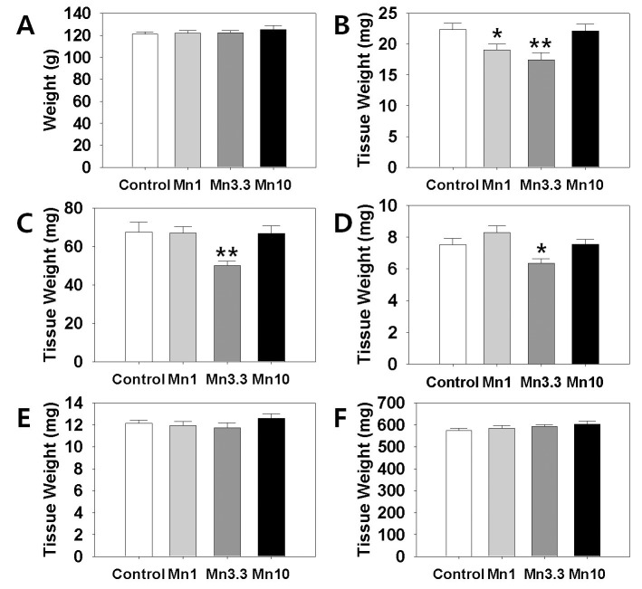
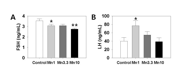
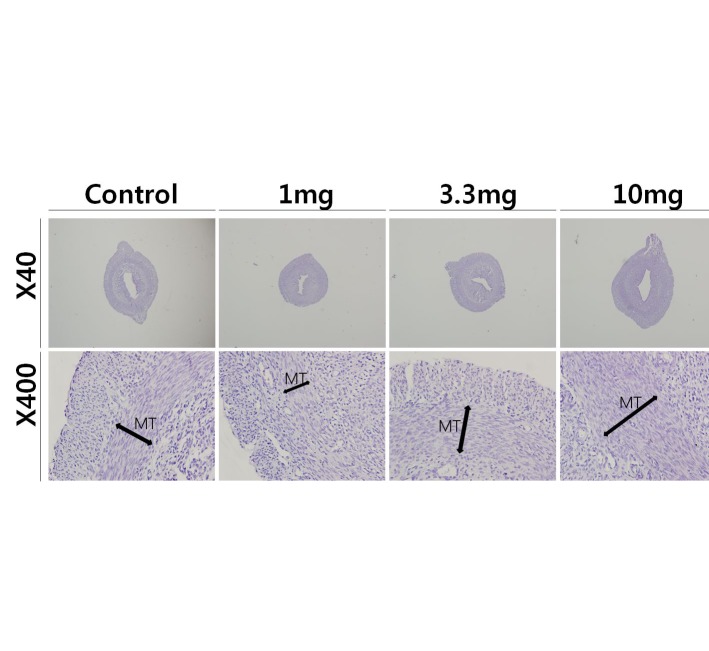
Manganese (Mn(2+)) is a trace element that is essential for normal physiology, and is predominantly obtained from food. Several lines of evidence, however, demonstrated that overexposure to MnCl2 exerts serious neurotoxicity, immunotoxicity and developmental toxicity, particularly in male. The present study aimed to evaluate the effect of 0, 1.0, 3.3, and 10 mg/kg/day doses of MnCl2 on the reproductive organs in the immature female rats. Rats (PND 22; S.D. strain) were exposed to MnCl2 (MnCl2 ∙ 4H2O) dissolved in drinking water for 2 weeks. The animals were sacrificed on PND 35, then the tissues were immediately removed and weighed. Histological studies were performed using the uteri tissue samples. Serum LH and FSH levels were measured with the specific ELISA kits. Body weights of the experimental group animals were not significantly different from those of control group animals. However, ovarian tissue weights in 1 mg and 3.3 mg MnCl2 dose groups were significantly lower than those of control animals (p<0.05 and p<0.01, respectively). Uterine tissue weights of 3.3 mg dose MnCl2 groups were significantly lower than those of control animals (p<0.01), while the 1 mg MnCl2 dose and 10 mg MnCl2 dose failed to induce any change in uterine weight. Similarly, only 3.3 mg MnCl2 dose could induce the significant decrease in the oviduct weight compared to the control group (p<0.05). Non-reproductive tissues such as adrenal and kidney failed to respond to all doses of MnCl2 exposure. The uterine histology revealed that the MnCl2 exposure could affect the myometrial cell proliferation particularly in 3.3 mg dose and 10mg dose group. Serum FSH levels were significantly decreased in 1mg MnCl2 dose and 10 MnCl2 mg groups (p<0.05 and p<0.01, respectively). In contrast, treatment with 1 mg MnCl2 dose induced a significant increment of serum LH level (p<0.05). The present study demonstrated that MnCl2 exposure is capable of inducing abnormal development of reproductive tissues, at least to some extent, and altered gonadotropin secretions in immature female rats. Combined with the well-defined actions of this metal on GnRH and prolactin secretion, one can suggest the Mn(2+) might be a potential environmental mediator which is involved in the female pubertal process.
锰(Mn(2+))是一种对正常生理功能至关重要的微量元素,主要从食物中获取。然而,多项证据表明,过度暴露于氯化锰会产生严重的神经毒性、免疫毒性和发育毒性,尤其是对雄性。本研究旨在评估0、1.0、3.3和10毫克/千克/天剂量的氯化锰对未成熟雌性大鼠生殖器官的影响。将大鼠(出生后第22天;SD品系)暴露于溶解在饮用水中的氯化锰(MnCl2 ∙ 4H2O)中2周。在出生后第35天处死动物,然后立即取出组织并称重。使用子宫组织样本进行组织学研究。用特定的ELISA试剂盒测量血清促黄体生成素(LH)和促卵泡生成素(FSH)水平。实验组动物的体重与对照组动物的体重无显著差异。然而,1毫克和3.3毫克氯化锰剂量组的卵巢组织重量显著低于对照组动物(分别为p<0.05和p<0.01)。3.3毫克剂量氯化锰组的子宫组织重量显著低于对照组动物(p<0.01),而1毫克氯化锰剂量组和10毫克氯化锰剂量组未能引起子宫重量的任何变化。同样,与对照组相比,只有3.3毫克氯化锰剂量可导致输卵管重量显著降低(p<0.05)。肾上腺和肾脏等非生殖组织对所有剂量的氯化锰暴露均无反应。子宫组织学显示,氯化锰暴露可影响子宫肌层细胞增殖,尤其是在3.3毫克剂量组和10毫克剂量组。1毫克氯化锰剂量组和10毫克氯化锰剂量组的血清FSH水平显著降低(分别为p<0.05和p<0.01)。相反,1毫克氯化锰剂量处理可导致血清LH水平显著升高(p<0.05)。本研究表明,氯化锰暴露至少在一定程度上能够诱导未成熟雌性大鼠生殖组织的异常发育,并改变促性腺激素的分泌。结合这种金属对促性腺激素释放激素(GnRH)和催乳素分泌的明确作用,可以推测Mn(2+)可能是参与雌性青春期过程的潜在环境介质。