Prince Daniel E, Greisberg Justin K
Memorial Sloan Kettering Cancer Center, 1275 York Avenue, Howard 1013, New York, NY, 10065, USA.
New York Presbyterian Hospital, Columbia University, New York, NY, USA.
J Orthop Traumatol. 2015 Dec;16(4):335-41. doi: 10.1007/s10195-015-0350-2. Epub 2015 May 10.
The primary goal of this study was to identify nitric oxide (NO)-induced apoptosis in traumatized chondrocytes in intra-articular lower extremity fractures and the secondary goal was to identify the timeline of NO-induced apoptosis after injury.
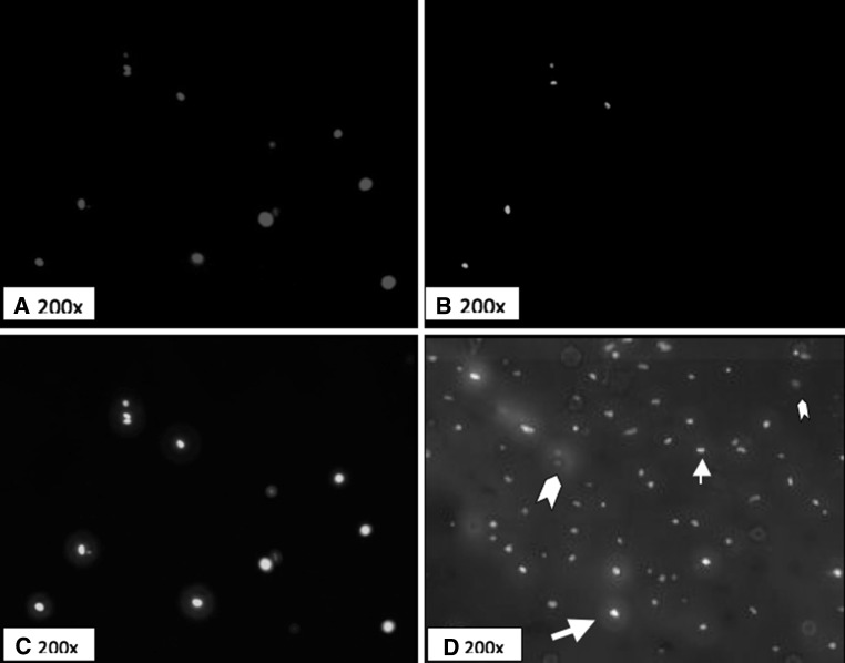
This is a prospective collection of samples of human cartilage harvested at the time of surgery to measure apoptotic cell death and the presence of NO by immunohistochemistry. Three patients met the criteria for control subjects and eight patients sustained high-energy intra-articular fractures and were included in the study. Subjects who sustained intra-articular acetabular, tibial, calcaneal and talus fracture had articular cartilage harvested at the time of surgical intervention. All 8 patients underwent open reduction and internal fixation of the displaced intra-articular fractures. The main outcome measures were rate of apoptosis, degree of NO-induced apoptosis in chondrocytes, and the timeline of NO-induced apoptosis after high-energy trauma.
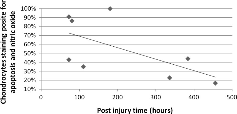
The percentage of apoptotic chondrocytes was higher in impacted samples than in normal cartilage (56 vs 4 %), confirming the presence of apoptosis after intra-articular fracture. The percentage of cells with NO was greater in apoptotic cells than in normal cells (59 vs 20 %), implicating NO-induction of apoptosis. The correlation between chondrocyte apoptosis and increasing time from injury was found to be -0.615, indicating a decreasing rate of apoptosis post injury.
The data showed the involvement of NO-induced apoptosis of chondrocytes after high-energy trauma, which decreased with time from injury.
本研究的主要目标是确定一氧化氮(NO)诱导的关节内下肢骨折创伤软骨细胞凋亡,次要目标是确定损伤后NO诱导凋亡的时间线。
这是一项前瞻性研究,收集手术时获取的人类软骨样本,通过免疫组织化学测量凋亡细胞死亡情况及NO的存在。3例患者符合对照标准,8例患者遭受高能关节内骨折并纳入研究。遭受髋臼、胫骨、跟骨和距骨关节内骨折的受试者在手术干预时采集关节软骨。所有8例患者均接受了移位关节内骨折的切开复位内固定术。主要观察指标为凋亡率、软骨细胞中NO诱导凋亡的程度以及高能创伤后NO诱导凋亡的时间线。
受冲击样本中凋亡软骨细胞的百分比高于正常软骨(56%对4%),证实关节内骨折后存在凋亡。凋亡细胞中含NO的细胞百分比高于正常细胞(59%对20%),提示NO诱导凋亡。发现软骨细胞凋亡与受伤时间增加之间的相关性为-0.615,表明损伤后凋亡率降低。
数据显示高能创伤后存在NO诱导的软骨细胞凋亡,且随受伤时间推移而降低。