• 文献检索
  • 文档翻译
  • 深度研究
  • 学术资讯
  • Suppr Zotero 插件Zotero 插件
  • 邀请有礼
  • 套餐&价格
  • 历史记录
应用&插件
Suppr Zotero 插件Zotero 插件浏览器插件Mac 客户端Windows 客户端微信小程序
定价
高级版会员购买积分包购买API积分包
服务
文献检索文档翻译深度研究API 文档MCP 服务
关于我们
关于 Suppr公司介绍联系我们用户协议隐私条款
关注我们

Suppr 超能文献

核心技术专利:CN118964589B侵权必究
粤ICP备2023148730 号-1Suppr @ 2026

文献检索

告别复杂PubMed语法,用中文像聊天一样搜索,搜遍4000万医学文献。AI智能推荐,让科研检索更轻松。

立即免费搜索

文件翻译

保留排版,准确专业,支持PDF/Word/PPT等文件格式,支持 12+语言互译。

免费翻译文档

深度研究

AI帮你快速写综述,25分钟生成高质量综述,智能提取关键信息,辅助科研写作。

立即免费体验

源自人主动脉原代培养的动脉粥样硬化及年龄相关的多核变异内皮细胞

Atherosclerosis- and age-related multinucleated variant endothelial cells in primary culture from human aorta.

作者信息

Tokunaga O, Fan J L, Watanabe T

机构信息

Department of Pathology, Saga Medical School, Nabeshima-Machi, Japan.

出版信息

Am J Pathol. 1989 Dec;135(6):967-76.

PMID:2596578
原文链接:https://pmc.ncbi.nlm.nih.gov/articles/PMC1880500/
Abstract

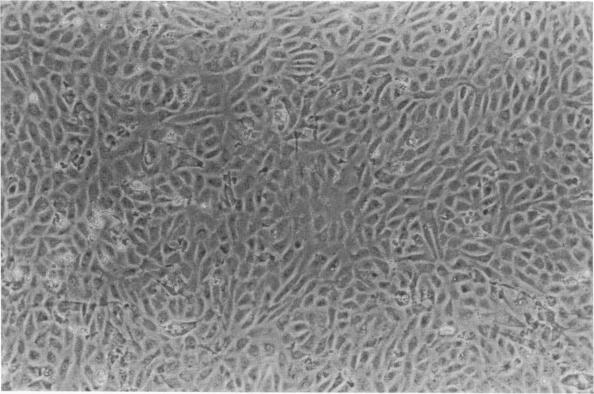
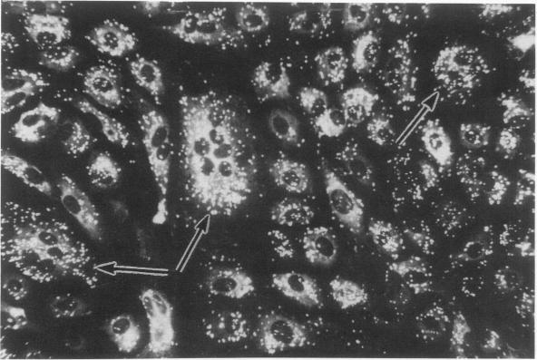
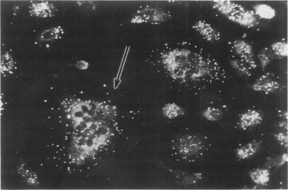
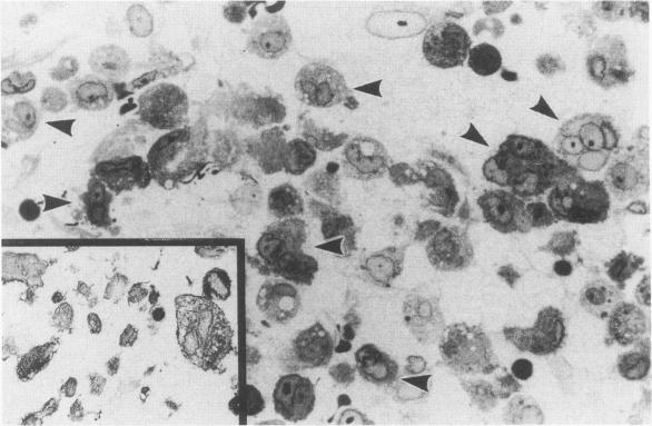
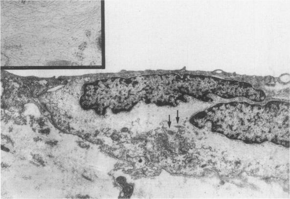
Endothelial cells were cultured from human aortas and inferior venae cavae of autopsied subjects ranging in age from infancy to 85 years. Endothelial cells in 32 of more than 100 attempted cultures were pure enough for evaluation. Emerged endothelial cells in primary culture were classified into two types: typical endothelium and variant endothelium. Typical endothelial cells were small, round to polygonal shaped, and were arranged uniformly. Their diameter ranged from 50 to 70 microns. Variant endothelial cells were larger, ranging from 100 to 200 microns in diameter, and giant endothelial cells measuring more than 250 microns in diameter were scattered among them. Variant endothelial cells were usually multinucleated and possessed endothelium-specific markers of vWF and Weibel-Palade bodies. No incorporation of [3H]thymidine was found in the nuclei of cultured variant endothelial cells. Although most cultured endothelial cells were of the typical type, variant endothelial cells were interspersed throughout the culture. The ratio of variant endothelial cells to typical cells correlated well with the severity of atherosclerosis, but less so with aging. The number of variant endothelial cells in cultures from inferior venae cavae was slight and constant throughout all age groups. The presence of multinucleated endothelial cells in in vivo aortas was confirmed by both scanning and transmission electron microscopy. They sometimes existed in colonies in the aortas from elderly subjects with intimal-thickened or advanced atherosclerotic lesions. These results indicate that variant endothelial cells were present in vivo and their ratio in primary culture reflected the in vivo population. It is likely that these cells were formed by adhesion of adjacent typical endothelial cells and that this process was affected more by atherosclerosis than by aging. Although it is not clear if the multinucleated variant cells were formed before the formation of atherosclerotic plaque or after the plaque formation, they will contribute to further development of atherosclerotic lesions, which in turn cause malfunction of the cell membrane. We suggest that there is a cyclic effect of these processes for multiplication of the variant endothelial cells and advancement of atherosclerotic lesions.

摘要

从年龄从婴儿期到85岁的尸检对象的人主动脉和下腔静脉中培养内皮细胞。在100多次尝试培养中,有32次培养出的内皮细胞纯度足以进行评估。原代培养中出现的内皮细胞分为两种类型:典型内皮细胞和变异内皮细胞。典型内皮细胞体积小,呈圆形至多边形,排列均匀。其直径范围为50至70微米。变异内皮细胞较大,直径范围为100至200微米,直径超过250微米的巨大内皮细胞散在于其中。变异内皮细胞通常为多核,并具有内皮特异性标志物vWF和魏-帕小体。在培养的变异内皮细胞核中未发现[3H]胸腺嘧啶核苷的掺入。虽然大多数培养的内皮细胞为典型类型,但变异内皮细胞散布于整个培养物中。变异内皮细胞与典型细胞的比例与动脉粥样硬化的严重程度密切相关,但与衰老的相关性较小。下腔静脉培养物中变异内皮细胞的数量在所有年龄组中都很少且恒定。通过扫描电子显微镜和透射电子显微镜均证实了体内主动脉中存在多核内皮细胞。它们有时存在于患有内膜增厚或晚期动脉粥样硬化病变的老年受试者主动脉中的菌落中。这些结果表明,变异内皮细胞存在于体内,其在原代培养中的比例反映了体内的细胞群体。这些细胞可能是由相邻的典型内皮细胞粘附形成的,并且这个过程受动脉粥样硬化的影响比受衰老的影响更大。虽然尚不清楚多核变异细胞是在动脉粥样硬化斑块形成之前还是之后形成的,但它们将有助于动脉粥样硬化病变的进一步发展,进而导致细胞膜功能障碍。我们认为这些过程对于变异内皮细胞的增殖和动脉粥样硬化病变的进展存在循环效应。

https://cdn.ncbi.nlm.nih.gov/pmc/blobs/db6d/1880500/9726b1ac956f/amjpathol00120-0021-a.jpg
https://cdn.ncbi.nlm.nih.gov/pmc/blobs/db6d/1880500/56d071e45f0e/amjpathol00120-0016-a.jpg
https://cdn.ncbi.nlm.nih.gov/pmc/blobs/db6d/1880500/8b42f4355ab6/amjpathol00120-0016-b.jpg
https://cdn.ncbi.nlm.nih.gov/pmc/blobs/db6d/1880500/7f2fb91035b8/amjpathol00120-0017-a.jpg
https://cdn.ncbi.nlm.nih.gov/pmc/blobs/db6d/1880500/1f206ddaf883/amjpathol00120-0017-b.jpg
https://cdn.ncbi.nlm.nih.gov/pmc/blobs/db6d/1880500/efcc39db4dbc/amjpathol00120-0018-a.jpg
https://cdn.ncbi.nlm.nih.gov/pmc/blobs/db6d/1880500/4154fe8f7c77/amjpathol00120-0018-b.jpg
https://cdn.ncbi.nlm.nih.gov/pmc/blobs/db6d/1880500/a67263bf304a/amjpathol00120-0019-a.jpg
https://cdn.ncbi.nlm.nih.gov/pmc/blobs/db6d/1880500/9726b1ac956f/amjpathol00120-0021-a.jpg

相似文献

1
Atherosclerosis- and age-related multinucleated variant endothelial cells in primary culture from human aorta.源自人主动脉原代培养的动脉粥样硬化及年龄相关的多核变异内皮细胞
Am J Pathol. 1989 Dec;135(6):967-76.
2
Endothelial cell heterogeneity in experimentally-induced rabbit atherosclerosis. Demonstration of multinucleated giant endothelial cells by scanning electron microscopy and cell culture.实验性诱导兔动脉粥样硬化中的内皮细胞异质性。通过扫描电子显微镜和细胞培养证实多核巨内皮细胞的存在。
Virchows Arch. 1994;425(5):521-9. doi: 10.1007/BF00197556.
3
Multinucleated variant endothelial cells (MVECs) in human aorta: chromosomal aneuploidy and elevated uptake of LDL.人主动脉中的多核变异内皮细胞(MVECs):染色体非整倍性与低密度脂蛋白摄取增加
Semin Thromb Hemost. 1998;24(3):279-84. doi: 10.1055/s-2007-995855.
4
Primary culture of endothelial cells from atherosclerotic human aorta. Part 1. Identification, morphological and ultrastructural characteristics of two endothelial cell subpopulations.来自动脉粥样硬化人主动脉的内皮细胞原代培养。第1部分。两种内皮细胞亚群的鉴定、形态学和超微结构特征。
Atherosclerosis. 1986 Jan;59(1):1-19. doi: 10.1016/0021-9150(86)90027-4.
5
Atherosclerosis and endothelium. Part 1. A simple method of endothelial cell culture from human atherosclerotic aorta.
Acta Pathol Jpn. 1987 Apr;37(4):527-36.
6
Multinucleated variant endothelial cells (MVECs) of human aorta: expression of tumor suppressor gene p53 and relationship to atherosclerosis and aging.人主动脉的多核变异内皮细胞(MVECs):肿瘤抑制基因p53的表达及其与动脉粥样硬化和衰老的关系
Endothelium. 1998;6(2):123-32. doi: 10.3109/10623329809072199.
7
Multinucleated variant endothelial cell. Its characterization and relation to atherosclerosis.多核变异内皮细胞。其特征及与动脉粥样硬化的关系。
Ann N Y Acad Sci. 1990;598:217-22. doi: 10.1111/j.1749-6632.1990.tb42293.x.
8
[Human aorta: endothelium and atheroma (author's transl)].[人类主动脉:内皮与动脉粥样硬化(作者译)]
Ann Pathol. 1981;1(4):271-9.
9
Activity of ATPase and 5'nucleotidase in endothelium of human atherosclerotic aortas.
Cor Vasa. 1992;34(3):238-45.
10
Formation of multinucleated variant endothelial cells in vitro and investigation of MVECs' features.体外多核变异内皮细胞的形成及多核内皮细胞特征的研究。
Fukuoka Igaku Zasshi. 1999 Oct;90(10):377-91.

引用本文的文献

1
Endothelial Senescence: From Macro- to Micro-Vasculature and Its Implications on Cardiovascular Health.内皮衰老:从大血管到微血管及其对心血管健康的影响。
Int J Mol Sci. 2024 Feb 6;25(4):1978. doi: 10.3390/ijms25041978.
2
Evaluating the antioxidant potential of resveratrol-gold nanoparticles in preventing oxidative stress in endothelium on a chip.评估白藜芦醇-金纳米粒子在预防芯片内皮氧化应激方面的抗氧化潜力。
Sci Rep. 2023 Dec 1;13(1):21344. doi: 10.1038/s41598-023-47291-6.
3
New Dawn for Atherosclerosis: Vascular Endothelial Cell Senescence and Death.

本文引用的文献

1
NEW CYTOPLASMIC COMPONENTS IN ARTERIAL ENDOTHELIA.动脉内皮细胞中的新细胞质成分
J Cell Biol. 1964 Oct;23(1):101-12. doi: 10.1083/jcb.23.1.101.
2
The human endothelial cell in tissue culture.组织培养中的人内皮细胞。
Z Zellforsch Mikrosk Anat. 1963;60:69-79. doi: 10.1007/BF00329383.
3
The quantitative appraisal of atherosclerosis.动脉粥样硬化的定量评估
动脉粥样硬化的新曙光:血管内皮细胞衰老与死亡。
Int J Mol Sci. 2023 Oct 13;24(20):15160. doi: 10.3390/ijms242015160.
4
An integrative review of nonobvious puzzles of cellular and molecular cardiooncology.细胞与分子心脏肿瘤学不明显难题的综合回顾。
Cell Mol Biol Lett. 2023 May 23;28(1):44. doi: 10.1186/s11658-023-00451-y.
5
The Role of Mitochondrial Mutations in Chronification of Inflammation: Hypothesis and Overview of Own Data.线粒体突变在炎症慢性化中的作用:假说及自身数据概述
Life (Basel). 2022 Jul 29;12(8):1153. doi: 10.3390/life12081153.
6
Large Multinucleated Variant Endothelial Cells in Allograft Kidney Microvasculature: A Biopsy Series.移植肾微血管中的大型多核变异内皮细胞:一项活检系列研究
Kidney Med. 2022 Jan 13;4(3):100411. doi: 10.1016/j.xkme.2022.100411. eCollection 2022 Mar.
7
Local Accumulation of Lymphocytes in the Intima of Human Aorta Is Associated with Giant Multinucleated Endothelial Cells: Possible Explanation for Mosaicism of Atherosclerosis.人类主动脉内皮下的淋巴细胞局部聚集与巨大多核内皮细胞有关:动脉粥样硬化镶嵌现象的可能解释。
Int J Mol Sci. 2022 Jan 19;23(3):1059. doi: 10.3390/ijms23031059.
8
Chronic irradiation of human cells reduces histone levels and deregulates gene expression.慢性辐射会降低人类细胞中的组蛋白水平,并使基因表达失调。
Sci Rep. 2020 Feb 10;10(1):2200. doi: 10.1038/s41598-020-59163-4.
9
Multinucleation of Incubated Cells and Their Morphological Differences Compared to Mononuclear Cells.培养细胞的多核化及其与单核细胞相比的形态学差异。
Micromachines (Basel). 2019 Feb 25;10(2):156. doi: 10.3390/mi10020156.
10
Pathology and biology of radiation-induced cardiac disease.放射性心脏病的病理学与生物学
J Radiat Res. 2016 Sep;57(5):439-448. doi: 10.1093/jrr/rrw064. Epub 2016 Jul 15.
Am J Pathol. 1957 Sep-Oct;33(5):875-85.
4
Reactions to injury of vascular endothelium with special reference to the problem of thrombosis.血管内皮损伤反应,特别涉及血栓形成问题。
J Pathol Bacteriol. 1955 Jan-Apr;69(1-2):283-93. doi: 10.1002/path.1700690136.
5
[Endothelium of aorta].[主动脉内皮]
Virchows Arch Pathol Anat Physiol Klin Med. 1952;322(6):662-94. doi: 10.1007/BF00957503.
6
Isolation and growth of endothelial cells from the microvessels of the newborn human foreskin in cell culture.在细胞培养中从新生儿包皮微血管分离内皮细胞并使其生长。
J Invest Dermatol. 1980 Oct;75(4):316-21. doi: 10.1111/1523-1747.ep12530941.
7
Cultured endothelial cells derived from the human iliac arteries.源自人髂动脉的培养内皮细胞。
In Vitro. 1982 Oct;18(10):859-66. doi: 10.1007/BF02796327.
8
Nitroglycerin stimulates synthesis of prostacyclin by cultured human endothelial cells.硝酸甘油可刺激培养的人内皮细胞合成前列环素。
J Clin Invest. 1981 Mar;67(3):762-9. doi: 10.1172/JCI110093.
9
Heterogeneity of endothelium in human aorta. A quantitative analysis by scanning electron microscopy.
Atherosclerosis. 1984 Jan;50(1):35-52. doi: 10.1016/0021-9150(84)90006-6.
10
Human endothelial cells: use of heparin in cloning and long-term serial cultivation.人内皮细胞:肝素在克隆和长期连续培养中的应用。
Science. 1983 Nov 11;222(4624):623-5. doi: 10.1126/science.6635659.