Saleem Azeem, Searle Graham E, Kenny Laura M, Huiban Mickael, Kozlowski Kasia, Waldman Adam D, Woodley Laura, Palmieri Carlo, Lowdell Charles, Kaneko Tomomi, Murphy Philip S, Lau Mike R, Aboagye Eric O, Coombes Raoul C
Imanova Centre for Imaging Sciences, Imperial College London, Hammersmith Hospital, Burlington Danes Building, Du Cane Road, London, W12 0NN, UK.
Department of Surgery and Cancer, Imperial College London, Charing Cross Hospital, Fulham Palace Road, London, W6 8RF, UK.
EJNMMI Res. 2015 Apr 30;5:30. doi: 10.1186/s13550-015-0103-5. eCollection 2015.
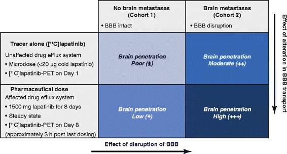
Brain metastases are common in human epidermal growth factor receptor (Her)-2-positive breast cancer. Drug access to brain metastases and normal brain is key to management of cranial disease. In this study, positron emission tomography (PET) scanning after administration of radiolabelled lapatinib was used to obtain direct evidence of cranial drug access.
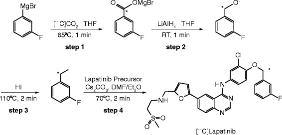
Patients with Her-2+ metastatic breast cancer either with at least one 1-cm diameter brain metastasis or without brain metastases underwent dynamic carbon-11 radiolabelled lapatinib ([(11)C]lapatinib)-PET. Less than 20 μg of [(11)C]lapatinib was administered before and after 8 days of oral lapatinib (1,500 mg once daily). Radial arterial blood sampling was performed throughout the 90-min scan. The contribution of blood volume activity to the tissue signal was excluded to calculate lapatinib uptake in normal brain and metastases. Partitioning of radioactivity between plasma and tissue (V T) was calculated and the tissue concentration of lapatinib derived. Plasma lapatinib levels were measured and adverse events noted.
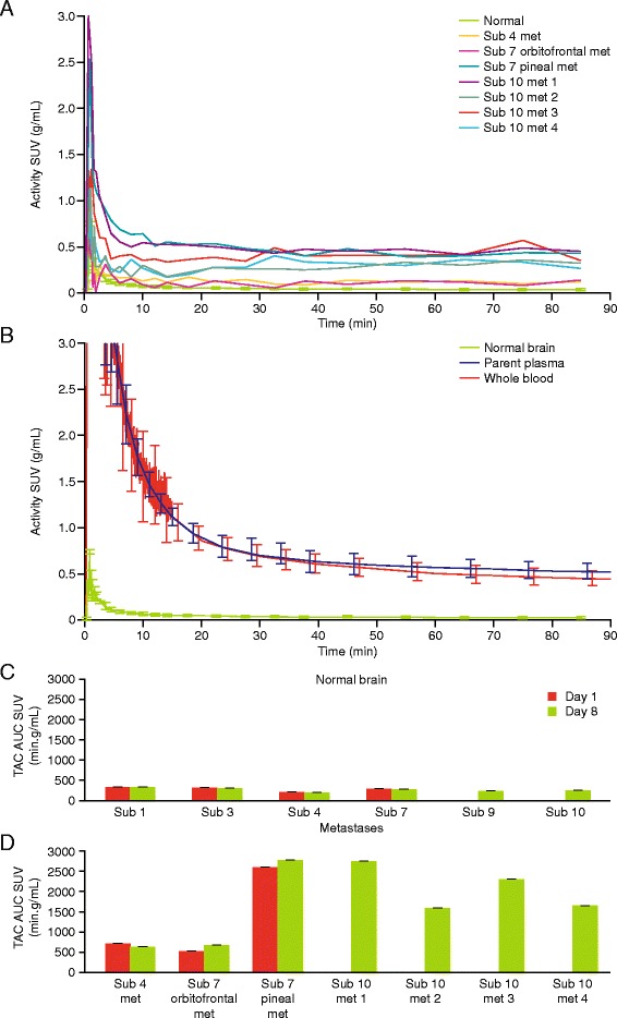
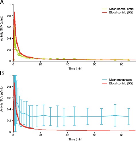
Six patients (three with brain metastases) were recruited. About 80% plasma radioactivity corresponded to intact [(11)C]lapatinib after 60 min. PET signal in the brain corresponded to circulating radioactivity levels, with no [(11)C]lapatinib uptake observed in normal brain tissue. In contrast, radioactivity uptake in cranial metastases was significantly higher (p = 0.002) than that could be accounted by circulating radioactivity levels, consistent with [(11)C]lapatinib uptake in brain metastases. There was no difference in lapatinib uptake between the baseline and day 8 scans, suggesting no effect of increased drug access by inhibition of the drug efflux proteins by therapeutic doses of lapatinib.
Increased lapatinib uptake was observed in brain metastases but not in normal brain.
ClinicalTrials.gov: NCT01290354.
脑转移在人表皮生长因子受体(Her)-2阳性乳腺癌中很常见。药物进入脑转移灶和正常脑组织是颅部疾病治疗的关键。在本研究中,给予放射性标记的拉帕替尼后进行正电子发射断层扫描(PET),以获取药物进入颅部的直接证据。
Her-2+转移性乳腺癌患者,至少有一个直径1厘米的脑转移灶或无脑转移灶,接受动态碳-11放射性标记拉帕替尼([(11)C]拉帕替尼)-PET检查。在口服拉帕替尼(1500毫克,每日一次)8天前后,给予少于20微克的[(11)C]拉帕替尼。在整个90分钟的扫描过程中进行桡动脉采血。排除血容量活性对组织信号的影响,以计算拉帕替尼在正常脑和转移灶中的摄取量。计算血浆与组织之间的放射性分配(VT),并得出拉帕替尼的组织浓度。测量血浆拉帕替尼水平并记录不良事件。
招募了6名患者(3名有脑转移灶)。60分钟后,约80%的血浆放射性对应完整的[(11)C]拉帕替尼。脑中的PET信号与循环放射性水平相对应,在正常脑组织中未观察到[(11)C]拉帕替尼的摄取。相比之下,颅部转移灶中的放射性摄取明显高于循环放射性水平所能解释的程度(p = 0.002),这与[(11)C]拉帕替尼在脑转移灶中的摄取一致。基线扫描和第8天扫描时拉帕替尼的摄取没有差异,这表明治疗剂量的拉帕替尼通过抑制药物外排蛋白增加药物进入没有效果。
在脑转移灶中观察到拉帕替尼摄取增加,但在正常脑中未观察到。
ClinicalTrials.gov:NCT01290354。