Pandit-Taskar Neeta, O'Donoghue Joseph A, Divgi Chaitanya R, Wills Eze A, Schwartz Lawrence, Gönen Mithat, Smith-Jones Peter, Bander Neil H, Scher Howard I, Larson Steven M, Morris Michael J
Molecular Imaging and Therapy Service, Department of Radiology, Memorial Sloan Kettering Cancer Center, 1275 York Avenue, New York, NY 10065 USA ; Department of Radiology, Weill Medical College of Cornell University, 1300 York Avenue, New York, NY 10065 USA.
Department of Medical Physics, Memorial Sloan Kettering Cancer Center, 1275 York Avenue, New York, NY 10065 USA.
EJNMMI Res. 2015 Apr 29;5:28. doi: 10.1186/s13550-015-0104-4. eCollection 2015.
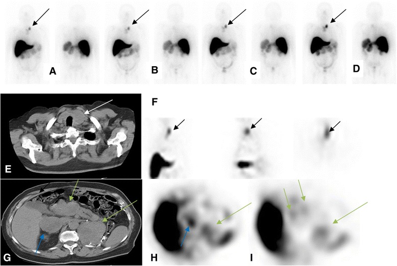
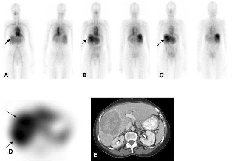
J591 is a monoclonal antibody that targets the external domain of the prostate-specific membrane antigen (PSMA). Besides prostate cancer cells, it also targets the neovasculature of non-prostate solid tumors. We provide an analysis of the antibody mass-dose dependency of lesion uptake and normal tissue retention, together with an assessment of lesion detectability using (111)In-J591 imaging, compared with conventional imaging in patients with a variety of solid tumors.
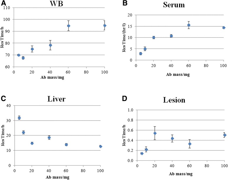
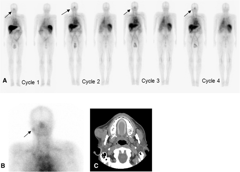
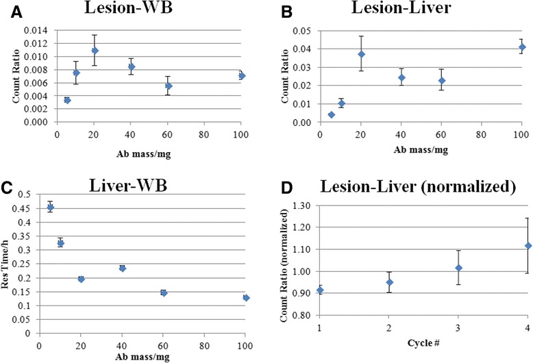
Twenty patients in six cohorts received fixed amounts (5, 10, 20, 40, 60, and 100 mg) of J591 in a phase I trial. A maximum of four administrations per patient was given, with each administration separated by 3 weeks. All antibody administrations included 370 MBq (10 mCi) of (111)In labeled to 2 mg of J591 via the chelating agent DOTA. Three whole body (WB) gamma camera scans with at least one SPECT scan, along with multiple WB count-rate measurements and blood samples, were obtained for all patients. The effect of escalating antibody mass on lesion uptake and normal tissue retention was evaluated using lesion, liver, serum, and WB residence times and ratios thereof for each treatment cycle. Lesion detectability using (111)In-J591 imaging was compared to the standard imaging on a lesion-by-lesion basis.
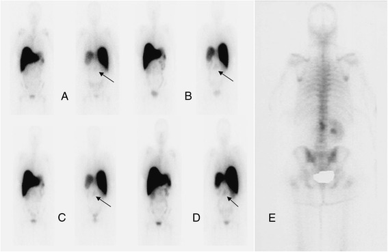
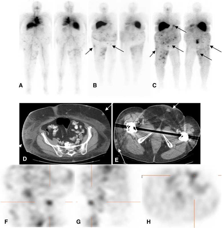
A total of 170 lesions in 20 patients were detected by standard or (111)In-J591 imaging. (111)In-J591 targeted both skeletal and soft tissue diseases in all tumor types. (111)In-J591 imaging identified 74% (20/27) of skeletal lesions, 53% (18/34) of nodes, and 64% (70/109) of other soft tissue/organ lesions. There was increasing (111)In-J591 uptake in lesions with increasing antibody mass-dose, coupled with decreasing retention in the liver for increments up to 20 mg, and no significant change at higher antibody mass.
Radiolabeled J591 antibody has potential as a targeting agent for solid tumor vasculature and lesion detection. Bone and soft tissue lesions arising from tumors of diverse origin were targeted by the anti-PSMA antibody J591. For the detection of lesions in these tumors by J591 antibody scans, an antibody mass of 20 mg is adequate. The optimal time of imaging is 5 to 7 days post-injection.
J591是一种单克隆抗体,靶向前列腺特异性膜抗原(PSMA)的胞外结构域。除前列腺癌细胞外,它还靶向非前列腺实体瘤的新生血管。我们分析了病变摄取和正常组织滞留的抗体质量剂量依赖性,并使用(111)In-J591成像评估病变的可检测性,与多种实体瘤患者的传统成像进行比较。
在一项I期试验中,六个队列的20名患者接受了固定剂量(5、10、20、40、60和mg)的J591。每位患者最多给药四次,每次给药间隔3周。所有抗体给药均包括通过螯合剂DOTA将370 MBq(10 mCi)的(111)In标记到2 mg J591上。为所有患者进行了三次全身(WB)γ相机扫描,至少一次SPECT扫描,以及多次WB计数率测量和血样采集。使用每个治疗周期的病变、肝脏、血清和WB滞留时间及其比率评估抗体质量增加对病变摄取和正常组织滞留的影响。将使用(111)In-J591成像的病变可检测性与标准成像在逐个病变的基础上进行比较。
通过标准或(111)In-J591成像在20名患者中总共检测到170个病变。(111)In-J591靶向所有肿瘤类型的骨骼和软组织疾病。(111)In-J591成像识别出74%(20/27)的骨骼病变、53%(18/34)的淋巴结和64%(70/109)的其他软组织/器官病变。随着抗体质量剂量的增加,病变中(111)In-J591的摄取增加,在抗体质量增加至20 mg时肝脏中的滞留减少,在更高抗体质量时无显著变化。
放射性标记的J591抗体有潜力作为实体瘤血管靶向剂和病变检测剂。抗PSMA抗体J591靶向多种来源肿瘤引起的骨骼和软组织病变。对于通过J591抗体扫描检测这些肿瘤中的病变,20 mg的抗体质量就足够了。最佳成像时间是注射后5至7天。