School of Health Sciences, Priority Research Centre for Physical Activity and Nutrition, University of Newcastle , Callaghan, NSW , Australia.
School of Health Sciences, Priority Research Centre for Translational Neuroscience and Mental Health, University of Newcastle , Callaghan, NSW , Australia.
Front Nutr. 2014 Jul 9;1:7. doi: 10.3389/fnut.2014.00007. eCollection 2014.
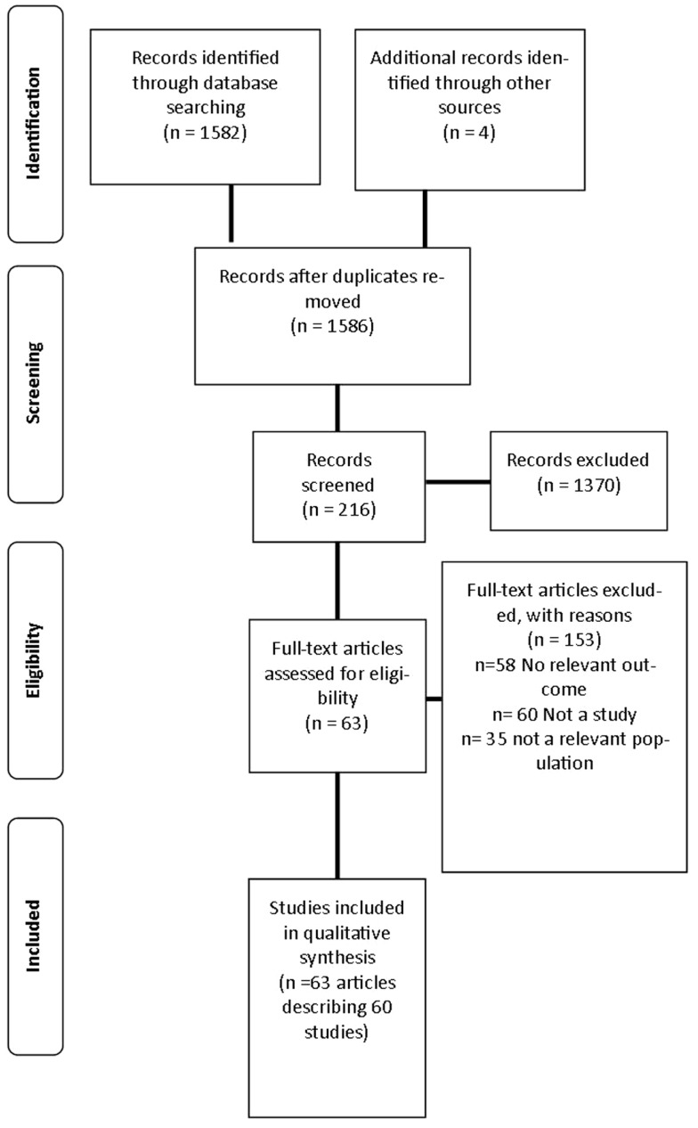
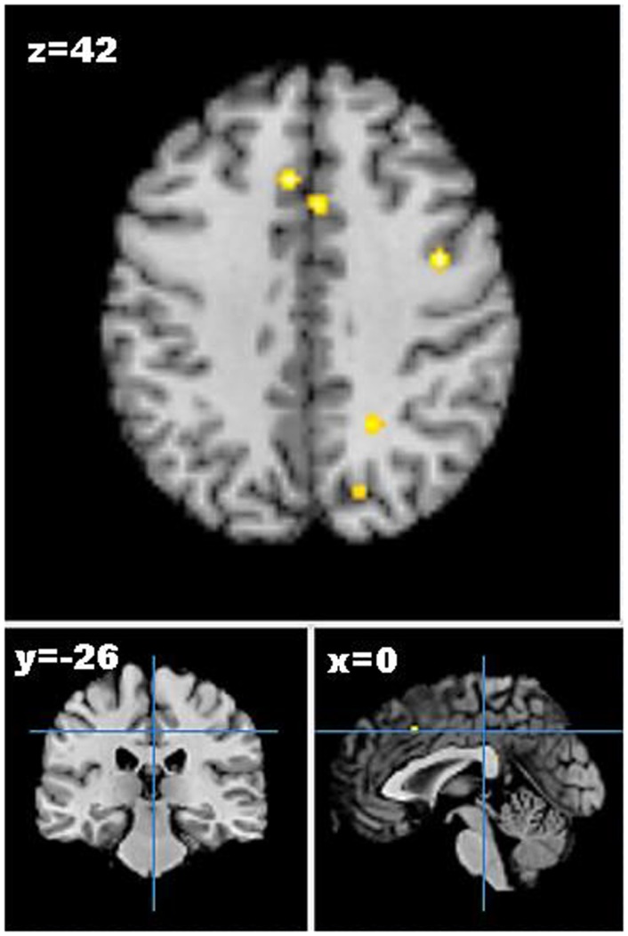
Emerging evidence from recent neuroimaging studies suggests that specific food-related behaviors contribute to the development of obesity. The aim of this review was to report the neural responses to visual food cues, as assessed by functional magnetic resonance imaging (fMRI), in humans of differing weight status. Published studies to 2014 were retrieved and included if they used visual food cues, studied humans >18 years old, reported weight status, and included fMRI outcomes. Sixty studies were identified that investigated the neural responses of healthy weight participants (n = 26), healthy weight compared to obese participants (n = 17), and weight-loss interventions (n = 12). High-calorie food images were used in the majority of studies (n = 36), however, image selection justification was only provided in 19 studies. Obese individuals had increased activation of reward-related brain areas including the insula and orbitofrontal cortex in response to visual food cues compared to healthy weight individuals, and this was particularly evident in response to energy dense cues. Additionally, obese individuals were more responsive to food images when satiated. Meta-analysis of changes in neural activation post-weight loss revealed small areas of convergence across studies in brain areas related to emotion, memory, and learning, including the cingulate gyrus, lentiform nucleus, and precuneus. Differential activation patterns to visual food cues were observed between obese, healthy weight, and weight-loss populations. Future studies require standardization of nutrition variables and fMRI outcomes to enable more direct comparisons between studies.
最近的神经影像学研究的新证据表明,特定的与食物相关的行为有助于肥胖的发展。本综述的目的是报告通过功能磁共振成像(fMRI)评估的不同体重状态的人类对视觉食物线索的神经反应。检索到截至 2014 年的已发表研究,如果它们使用视觉食物线索,研究 18 岁以上的人类,报告体重状况,并包括 fMRI 结果,则将其包括在内。确定了 60 项研究,研究了健康体重参与者(n=26),健康体重与肥胖参与者(n=17)和减肥干预措施(n=12)的神经反应。在大多数研究中(n=36)使用了高热量食物图像,但只有 19 项研究提供了图像选择的理由。与健康体重个体相比,肥胖个体对视觉食物线索的反应中,奖励相关大脑区域(包括岛叶和眶额皮质)的激活增加,而对于能量密集的线索则更为明显。此外,肥胖个体在饱腹感时对食物图像的反应更敏感。减肥后神经激活变化的荟萃分析显示,与情绪,记忆和学习相关的大脑区域(包括扣带回,豆状核和楔前叶)的研究中存在小面积的收敛。在肥胖,健康体重和减肥人群之间观察到对视觉食物线索的不同激活模式。未来的研究需要对营养变量和 fMRI 结果进行标准化,以能够在研究之间进行更直接的比较。