São Paulo Clínicas Liver Cancer Group, São Paulo, Brazil.
Clinics (Sao Paulo). 2015 Mar;70(3):207-13. doi: 10.6061/clinics/2015(03)10. Epub 2015 Mar 1.
Fibrolamellar hepatocellular carcinoma is a rare primary malignant liver tumor that differs from conventional hepatocellular carcinoma in several aspects. The aim of this study was to describe the clinical, surgical and histopathological features of fibrolamellar hepatocellular carcinoma and to analyze the factors associated with survival.
We identified 21 patients with histopathologically diagnosed fibrolamellar hepatocellular carcinoma over a 22-year period. Clinical information was collected from medical records and biopsies, and surgical specimens were reviewed.
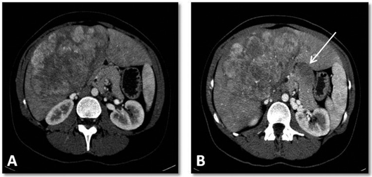
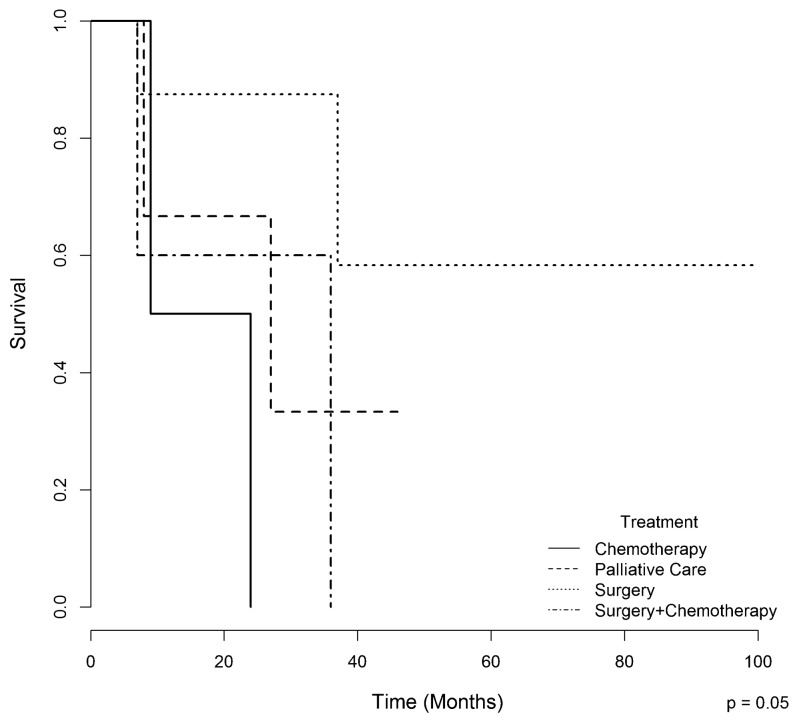
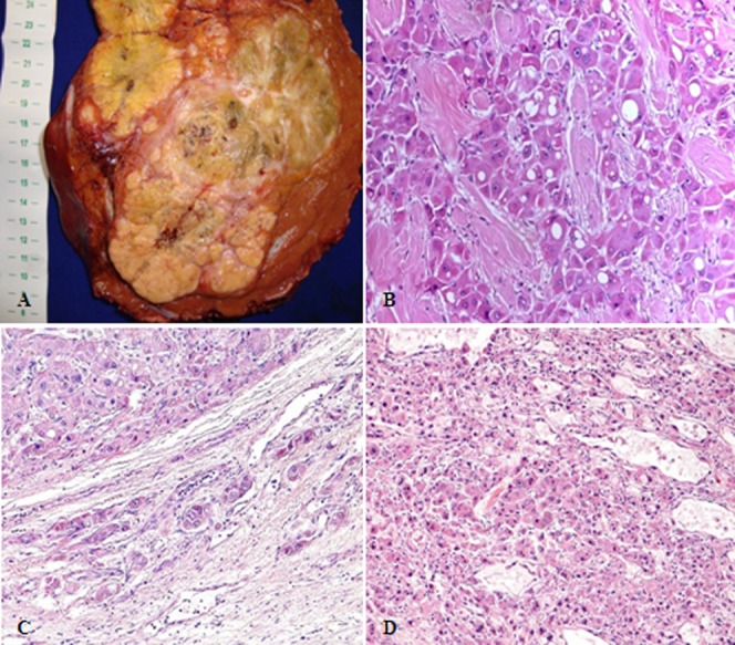
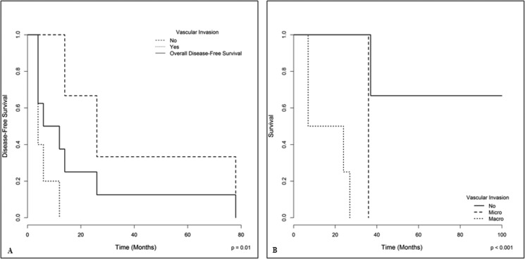
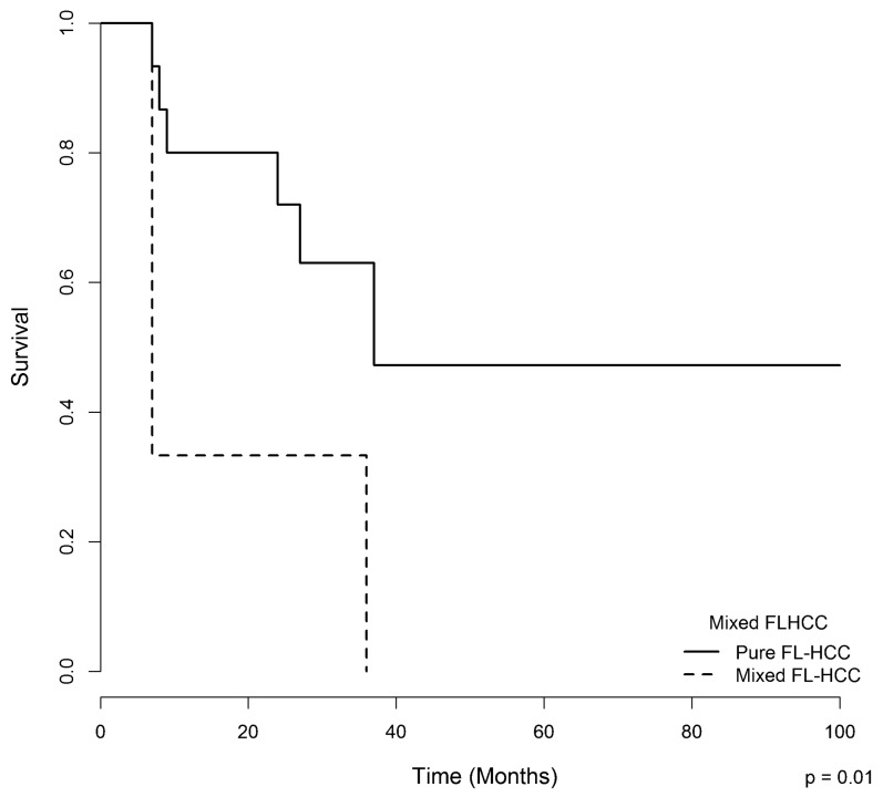
The median age at diagnosis was 20 years. Most patients were female (67%) and did not have associated chronic liver disease. Most patients had a single nodule, and the median tumor size was 120 mm. Vascular invasion was present in 31% of patients, and extra-hepatic metastases were present in 53%. Fourteen patients underwent surgery as the first-line therapy, three received chemotherapy, and four received palliative care. Eighteen patients had "pure fibrolamellar hepatocellular carcinoma," whereas three had a distinct area of conventional hepatocellular carcinoma and were classified as having "mixed fibrolamellar hepatocellular carcinoma." The median overall survival was 36 months. The presence of "mixed fibrolamellar hepatocellular carcinoma" and macrovascular invasion were predictors of poor survival. Vascular invasion was associated with an increased risk of recurrence in patients who underwent surgery.
Fibrolamellar hepatocellular carcinoma was more common in young female patients without chronic liver disease. Surgery was the first therapeutic option to achieve disease control, even in advanced cases. Vascular invasion was a risk factor for tumor recurrence. The presence of macrovascular invasion and areas of conventional hepatocellular carcinoma were directly related to poor survival.
纤维板层肝细胞癌是一种罕见的原发性肝脏恶性肿瘤,在多个方面与传统肝细胞癌不同。本研究旨在描述纤维板层肝细胞癌的临床、外科和组织病理学特征,并分析与生存相关的因素。
我们在 22 年的时间里确定了 21 例经组织病理学诊断为纤维板层肝细胞癌的患者。从病历和活检中收集临床信息,并对手术标本进行了回顾。
诊断时的中位年龄为 20 岁。大多数患者为女性(67%),且无相关慢性肝病。大多数患者有单个结节,肿瘤大小中位数为 120mm。31%的患者存在血管侵犯,53%的患者存在肝外转移。14 例患者作为一线治疗接受了手术,3 例接受了化疗,4 例接受了姑息治疗。18 例患者为“纯纤维板层肝细胞癌”,3 例为“混合纤维板层肝细胞癌”,有明显的传统肝细胞癌区域。中位总生存期为 36 个月。“混合纤维板层肝细胞癌”和大血管侵犯是生存不良的预测因素。血管侵犯与手术患者肿瘤复发风险增加相关。
纤维板层肝细胞癌在无慢性肝病的年轻女性中更为常见。手术是实现疾病控制的首选治疗方法,即使在晚期病例中也是如此。血管侵犯是肿瘤复发的危险因素。大血管侵犯和传统肝细胞癌区域的存在与生存不良直接相关。