Liu Zhigang, Patil Ishan Y, Jiang Tianyi, Sancheti Harsh, Walsh John P, Stiles Bangyan L, Yin Fei, Cadenas Enrique
Pharmacology & Pharmaceutical Sciences, School of Pharmacy, University of Southern California, Los Angeles, CA, 90089, United States of America; College of Food Science and Engineering, Northwest A&F University, Yangling, China.
Pharmacology & Pharmaceutical Sciences, School of Pharmacy, University of Southern California, Los Angeles, CA, 90089, United States of America.
PLoS One. 2015 May 29;10(5):e0128274. doi: 10.1371/journal.pone.0128274. eCollection 2015.
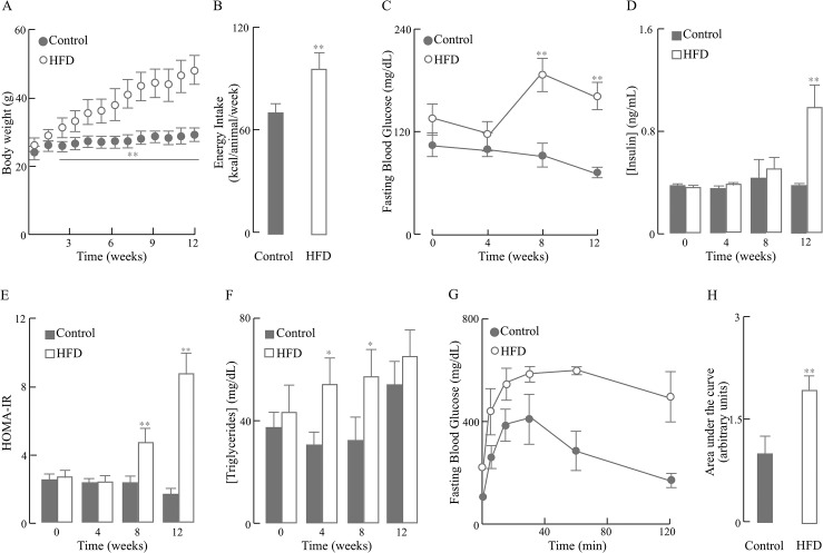
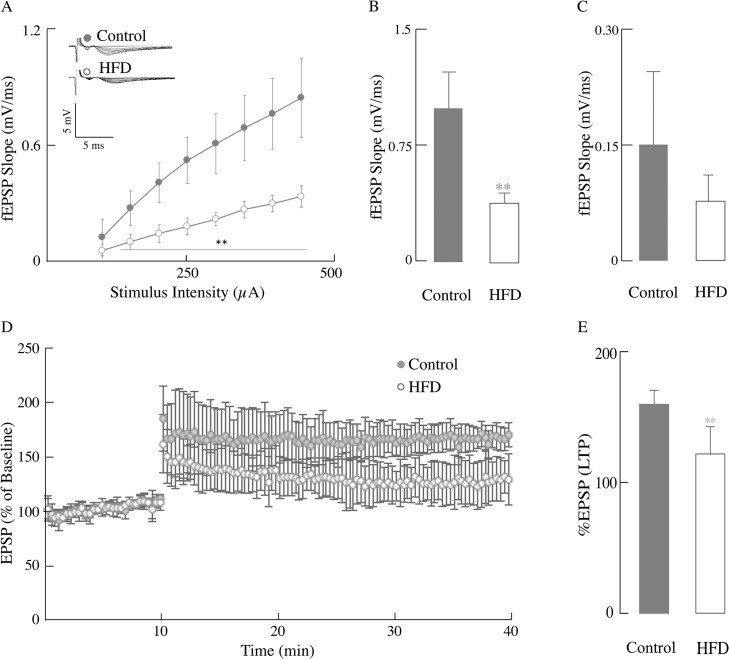
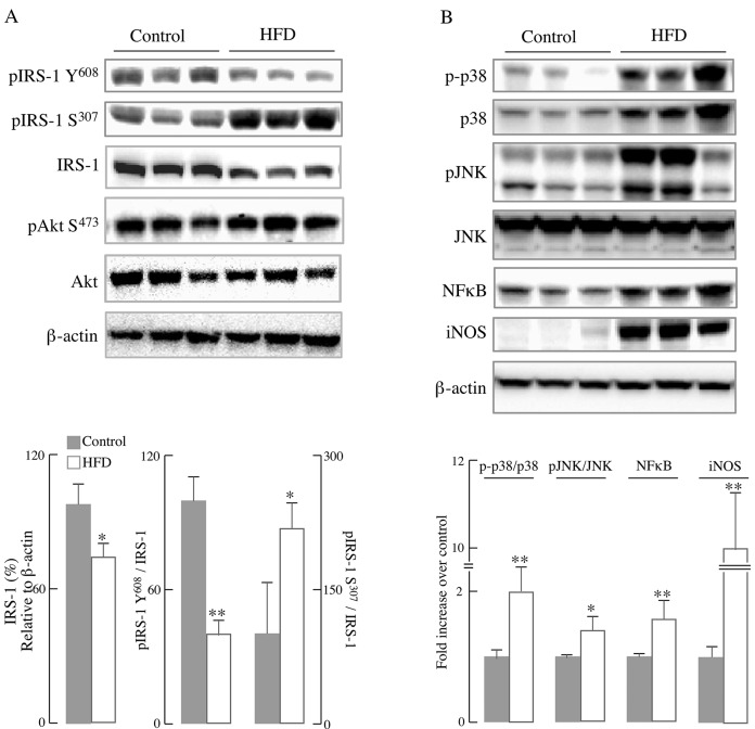
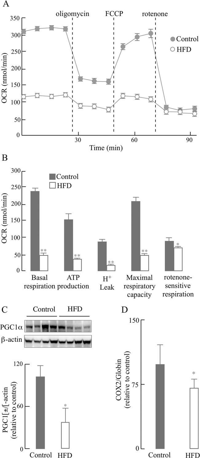
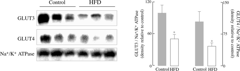
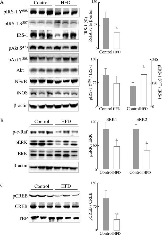
High-fat diet (HFD)-induced obesity is associated with insulin resistance, which may affect brain synaptic plasticity through impairment of insulin-sensitive processes underlying neuronal survival, learning, and memory. The experimental model consisted of 3 month-old C57BL/6J mice fed either a normal chow diet (control group) or a HFD (60% of calorie from fat; HFD group) for 12 weeks. This model was characterized as a function of time in terms of body weight, fasting blood glucose and insulin levels, HOMA-IR values, and plasma triglycerides. IRS-1/Akt pathway was assessed in primary hepatocytes and brain homogenates. The effect of HFD in brain was assessed by electrophysiology, input/output responses and long-term potentiation. HFD-fed mice exhibited a significant increase in body weight, higher fasting glucose- and insulin levels in plasma, lower glucose tolerance, and higher HOMA-IR values. In liver, HFD elicited (a) a significant decrease of insulin receptor substrate (IRS-1) phosphorylation on Tyr608 and increase of Ser307 phosphorylation, indicative of IRS-1 inactivation; (b) these changes were accompanied by inflammatory responses in terms of increases in the expression of NFκB and iNOS and activation of the MAP kinases p38 and JNK; (c) primary hepatocytes from mice fed a HFD showed decreased cellular oxygen consumption rates (indicative of mitochondrial functional impairment); this can be ascribed partly to a decreased expression of PGC1α and mitochondrial biogenesis. In brain, HFD feeding elicited (a) an inactivation of the IRS-1 and, consequentially, (b) a decreased expression and plasma membrane localization of the insulin-sensitive neuronal glucose transporters GLUT3/GLUT4; (c) a suppression of the ERK/CREB pathway, and (d) a substantial decrease in long-term potentiation in the CA1 region of hippocampus (indicative of impaired synaptic plasticity). It may be surmised that 12 weeks fed with HFD induce a systemic insulin resistance that impacts profoundly on brain activity, i.e., synaptic plasticity.
高脂饮食(HFD)诱导的肥胖与胰岛素抵抗有关,胰岛素抵抗可能通过损害神经元存活、学习和记忆所依赖的胰岛素敏感过程来影响脑突触可塑性。实验模型由3月龄的C57BL/6J小鼠组成,这些小鼠被喂食正常饲料(对照组)或高脂饮食(脂肪提供60%的热量;高脂饮食组)12周。该模型根据体重、空腹血糖和胰岛素水平、HOMA-IR值以及血浆甘油三酯随时间的变化情况进行表征。在原代肝细胞和脑匀浆中评估IRS-1/Akt信号通路。通过电生理学、输入/输出反应和长时程增强来评估高脂饮食对脑的影响。喂食高脂饮食的小鼠体重显著增加,血浆中空腹葡萄糖和胰岛素水平升高,葡萄糖耐量降低,HOMA-IR值升高。在肝脏中,高脂饮食导致:(a)胰岛素受体底物(IRS-1)酪氨酸608位点磷酸化显著降低,丝氨酸307位点磷酸化增加,表明IRS-1失活;(b)这些变化伴随着NFκB和iNOS表达增加以及MAP激酶p38和JNK激活所介导的炎症反应;(c)喂食高脂饮食的小鼠原代肝细胞显示细胞耗氧率降低(表明线粒体功能受损);这部分可归因于PGC1α表达降低和线粒体生物发生减少。在脑中,喂食高脂饮食导致:(a)IRS-1失活,进而(b)胰岛素敏感的神经元葡萄糖转运体GLUT3/GLUT4的表达和质膜定位降低;(c)ERK/CREB信号通路受到抑制,以及(d)海马体CA1区的长时程增强显著降低(表明突触可塑性受损)。可以推测,喂食12周高脂饮食会诱导全身性胰岛素抵抗,这对脑活动即突触可塑性产生深远影响。