• 文献检索
  • 文档翻译
  • 深度研究
  • 学术资讯
  • Suppr Zotero 插件Zotero 插件
  • 邀请有礼
  • 套餐&价格
  • 历史记录
应用&插件
Suppr Zotero 插件Zotero 插件浏览器插件Mac 客户端Windows 客户端微信小程序
定价
高级版会员购买积分包购买API积分包
服务
文献检索文档翻译深度研究API 文档MCP 服务
关于我们
关于 Suppr公司介绍联系我们用户协议隐私条款
关注我们

Suppr 超能文献

核心技术专利:CN118964589B侵权必究
粤ICP备2023148730 号-1Suppr @ 2026

文献检索

告别复杂PubMed语法,用中文像聊天一样搜索,搜遍4000万医学文献。AI智能推荐,让科研检索更轻松。

立即免费搜索

文件翻译

保留排版,准确专业,支持PDF/Word/PPT等文件格式,支持 12+语言互译。

免费翻译文档

深度研究

AI帮你快速写综述,25分钟生成高质量综述,智能提取关键信息,辅助科研写作。

立即免费体验

神经元缺氧会破坏线粒体融合。

Neuronal hypoxia disrupts mitochondrial fusion.

作者信息

Sanderson T H, Raghunayakula S, Kumar R

机构信息

Department of Emergency Medicine, Wayne State University School of Medicine, 540 E. Canfield, Detroit, MI, USA; Cardiovascular Research Institute, Wayne State University School of Medicine, 421E. Canfield, Detroit, MI, USA.

Department of Emergency Medicine, Wayne State University School of Medicine, 540 E. Canfield, Detroit, MI, USA.

出版信息

Neuroscience. 2015 Aug 20;301:71-8. doi: 10.1016/j.neuroscience.2015.05.078. Epub 2015 Jun 3.

DOI:10.1016/j.neuroscience.2015.05.078
PMID:26049142
原文链接:https://pmc.ncbi.nlm.nih.gov/articles/PMC4504771/
Abstract

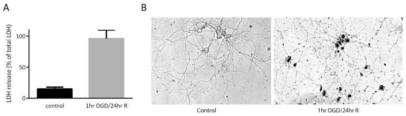
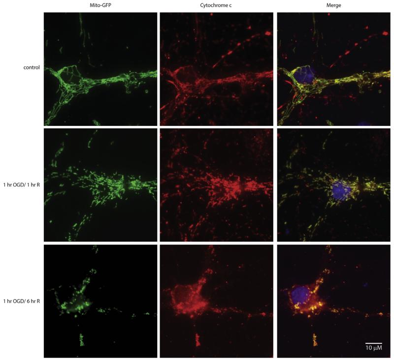
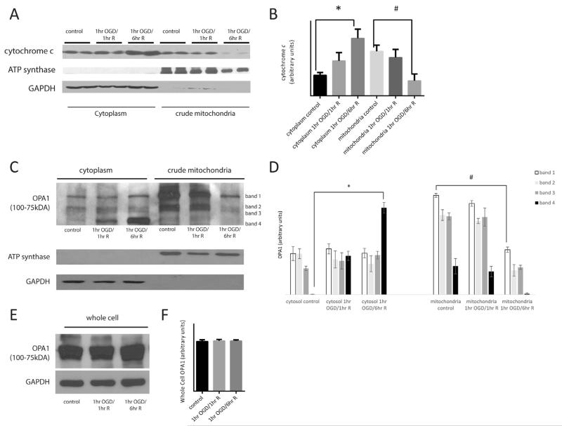
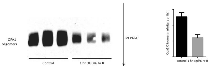
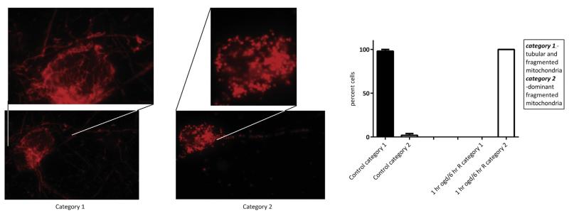
Brain ischemia/reperfusion injury results in death of vulnerable neurons and extensive brain damage. It is well known that mitochondrial release of cytochrome c (cyto c) is a hallmark of neuronal death, however the molecular events underlying this release are largely unknown. We tested the hypothesis that cyto c release is regulated by breakdown of the cristae architecture maintenance protein, optic atrophy 1 (OPA1), located in the inner mitochondrial membrane. We simulated ischemia/reperfusion in isolated primary rat neurons and interrogated OPA1 release from the mitochondria, OPA1 oligomeric breakdown, and concomitant dysfunction of mitochondrial dynamic state. We found that ischemia/reperfusion induces cyto c release and cell death that corresponds to multiple changes in OPA1, including: (i) translocation of the mitochondrial fusion protein OPA1 from the mitochondria to the cytosol, (ii) increase in the short isoform of OPA1, suggestive of proteolytic processing, (iii) breakdown of OPA1 oligomers in the mitochondria, and (iv) increased mitochondrial fission. Thus, we present novel evidence of a connection between release of cyto c from mitochondria and disruption of the mitochondrial fusion.

摘要

脑缺血/再灌注损伤会导致易损神经元死亡和广泛的脑损伤。众所周知,细胞色素c(cyto c)从线粒体释放是神经元死亡的一个标志,然而,这种释放背后的分子事件在很大程度上尚不清楚。我们检验了这样一个假设,即cyto c的释放受位于线粒体内膜的嵴结构维持蛋白视神经萎缩蛋白1(OPA1)的分解调控。我们在分离的原代大鼠神经元中模拟缺血/再灌注,并研究OPA1从线粒体的释放、OPA1寡聚体的分解以及伴随的线粒体动态状态功能障碍。我们发现,缺血/再灌注诱导cyto c释放和细胞死亡,这与OPA1的多种变化相对应,包括:(i)线粒体融合蛋白OPA1从线粒体转运至胞质溶胶;(ii)OPA1短异构体增加,提示存在蛋白水解过程;(iii)线粒体中OPA1寡聚体的分解;以及(iv)线粒体裂变增加。因此,我们提供了线粒体中cyto c释放与线粒体融合破坏之间存在联系的新证据。

https://cdn.ncbi.nlm.nih.gov/pmc/blobs/7ec1/4504771/26d53a7c7053/nihms-702430-f0005.jpg
https://cdn.ncbi.nlm.nih.gov/pmc/blobs/7ec1/4504771/68362b7fc12d/nihms-702430-f0001.jpg
https://cdn.ncbi.nlm.nih.gov/pmc/blobs/7ec1/4504771/a11f9393c64e/nihms-702430-f0002.jpg
https://cdn.ncbi.nlm.nih.gov/pmc/blobs/7ec1/4504771/d1cb29f5f8c9/nihms-702430-f0003.jpg
https://cdn.ncbi.nlm.nih.gov/pmc/blobs/7ec1/4504771/6cd2e20332d5/nihms-702430-f0004.jpg
https://cdn.ncbi.nlm.nih.gov/pmc/blobs/7ec1/4504771/26d53a7c7053/nihms-702430-f0005.jpg

相似文献

1
Neuronal hypoxia disrupts mitochondrial fusion.神经元缺氧会破坏线粒体融合。
Neuroscience. 2015 Aug 20;301:71-8. doi: 10.1016/j.neuroscience.2015.05.078. Epub 2015 Jun 3.
2
Mitochondrial dynamics following global cerebral ischemia.全脑缺血后的线粒体动力学
Mol Cell Neurosci. 2016 Oct;76:68-75. doi: 10.1016/j.mcn.2016.08.010. Epub 2016 Aug 25.
3
OPA1 overexpression ameliorates mitochondrial cristae remodeling, mitochondrial dysfunction, and neuronal apoptosis in prion diseases.OPA1 过表达改善朊病毒病中线粒体嵴重塑、线粒体功能障碍和神经元凋亡。
Cell Death Dis. 2019 Sep 24;10(10):710. doi: 10.1038/s41419-019-1953-y.
4
Optic atrophy 1 mediates mitochondria remodeling and dopaminergic neurodegeneration linked to complex I deficiency.视神经萎缩 1 介导与复合物 I 缺陷相关的线粒体重构和多巴胺能神经退行性变。
Cell Death Differ. 2013 Jan;20(1):77-85. doi: 10.1038/cdd.2012.95. Epub 2012 Aug 3.
5
Oxidization of optic atrophy 1 cysteines occurs during heart ischemia-reperfusion and amplifies cell death by oxidative stress.视萎缩 1 半胱氨酸的氧化发生在心脏缺血再灌注期间,并通过氧化应激放大细胞死亡。
Redox Biol. 2023 Jul;63:102755. doi: 10.1016/j.redox.2023.102755. Epub 2023 May 19.
6
N-terminal cleavage of the mitochondrial fusion GTPase OPA1 occurs via a caspase-independent mechanism in cerebellar granule neurons exposed to oxidative or nitrosative stress.线粒体融合 GTP 酶 OPA1 的 N 端切割发生在小脑颗粒神经元暴露于氧化或硝化应激时通过一种不依赖于胱天蛋白酶的机制。
Brain Res. 2013 Feb 4;1494:28-43. doi: 10.1016/j.brainres.2012.12.001. Epub 2012 Dec 7.
7
Mechanistic study of optic atrophy 1 in ischemia-reperfusion disease.对缺血再灌注疾病中 1 型视神经萎缩的机制研究。
J Mol Med (Berl). 2023 Feb;101(1-2):1-8. doi: 10.1007/s00109-022-02271-7. Epub 2022 Nov 23.
8
Lycium Barbarum Polysaccharides Decrease Hyperglycemia-Aggravated Ischemic Brain Injury through Maintaining Mitochondrial Fission and Fusion Balance.枸杞多糖通过维持线粒体分裂和融合平衡来降低高血糖加重的缺血性脑损伤。
Int J Biol Sci. 2017 Jul 7;13(7):901-910. doi: 10.7150/ijbs.18404. eCollection 2017.
9
Elevated hydrostatic pressure triggers release of OPA1 and cytochrome C, and induces apoptotic cell death in differentiated RGC-5 cells.升高的流体静压触发OPA1和细胞色素C的释放,并诱导分化的RGC-5细胞发生凋亡性细胞死亡。
Mol Vis. 2009;15:120-34. Epub 2009 Jan 19.
10
OPA1 processing in cell death and disease - the long and short of it.细胞死亡与疾病中的OPA1加工——其来龙去脉
J Cell Sci. 2016 Jun 15;129(12):2297-306. doi: 10.1242/jcs.159186. Epub 2016 May 17.

引用本文的文献

1
Analysis of neuronal cardiolipin and monolysocardiolipin from biological samples with cyclic ion mobility mass spectrometry.采用循环离子淌度质谱法分析生物样品中的神经元心磷脂和单溶血心磷脂。
Front Physiol. 2025 May 29;16:1592008. doi: 10.3389/fphys.2025.1592008. eCollection 2025.
2
Autophagy: a double-edged sword in ischemia-reperfusion injury.自噬:缺血再灌注损伤中的双刃剑
Cell Mol Biol Lett. 2025 Apr 7;30(1):42. doi: 10.1186/s11658-025-00713-x.
3
Decoding ischemic stroke: Perspectives on the endoplasmic reticulum, mitochondria, and their crosstalk.

本文引用的文献

1
Release of mitochondrial Opa1 following oxidative stress in HT22 cells.氧化应激后HT22细胞中线粒体Opa1的释放。
Mol Cell Neurosci. 2015 Jan;64:116-22. doi: 10.1016/j.mcn.2014.12.007. Epub 2015 Jan 8.
2
Mitochondrial dynamics: cell-type and hippocampal region specific changes following global cerebral ischemia.线粒体动力学:全脑缺血后细胞类型和海马区域的特异性变化
J Bioenerg Biomembr. 2015 Apr;47(1-2):13-31. doi: 10.1007/s10863-014-9575-7. Epub 2014 Sep 24.
3
Deregulation of mitochondria-shaping proteins Opa-1 and Drp-1 in manganese-induced apoptosis.
解读缺血性中风:内质网、线粒体及其相互作用的研究视角
Redox Biol. 2025 May;82:103622. doi: 10.1016/j.redox.2025.103622. Epub 2025 Mar 27.
4
Calycosin‑7‑O‑β‑D‑glucoside downregulates mitophagy by mitigating mitochondrial fission to protect HT22 cells from oxygen‑glucose deprivation/reperfusion‑induced injury.毛蕊异黄酮-7-O-β-D-葡萄糖苷通过减轻线粒体分裂来下调线粒体自噬,从而保护HT22细胞免受氧糖剥夺/再灌注诱导的损伤。
Mol Med Rep. 2025 Mar;31(3). doi: 10.3892/mmr.2025.13436. Epub 2025 Jan 17.
5
WTAP-mediated mA modification of TRIM22 promotes diabetic nephropathy by inducing mitochondrial dysfunction via ubiquitination of OPA1.WTAP 介导的 TRIM22 的 mA 修饰通过泛素化 OPA1 诱导线粒体功能障碍促进糖尿病肾病。
Redox Rep. 2024 Dec;29(1):2404794. doi: 10.1080/13510002.2024.2404794. Epub 2024 Sep 23.
6
Mitochondrial membrane potential and oxidative stress interact to regulate Oma1-dependent processing of Opa1 and mitochondrial dynamics.线粒体膜电位和氧化应激相互作用调节 Oma1 依赖的 Opa1 加工和线粒体动力学。
FASEB J. 2024 Sep 30;38(18):e70066. doi: 10.1096/fj.202400313R.
7
Leflunomide Treatment Does Not Protect Neural Cells following Oxygen-Glucose Deprivation (OGD) In Vitro.来氟米特治疗不能防止体外氧葡萄糖剥夺(OGD)后神经细胞的损伤。
Cells. 2024 Apr 4;13(7):631. doi: 10.3390/cells13070631.
8
Succinate-mediated reactive oxygen species production in the anoxia-tolerant epaulette () and grey carpet () sharks.琥珀酸介导的耐缺氧()和灰地毯()鲨鱼中的活性氧物质的产生。
Biol Lett. 2023 Oct;19(10):20230344. doi: 10.1098/rsbl.2023.0344. Epub 2023 Oct 11.
9
EGCG Attenuates CA1 Neuronal Death by Regulating GPx1, NF-κB S536 Phosphorylation and Mitochondrial Dynamics in the Rat Hippocampus following Status Epilepticus.表没食子儿茶素没食子酸酯通过调节癫痫持续状态后大鼠海马中谷胱甘肽过氧化物酶1、核因子κB S536磷酸化及线粒体动力学来减轻CA1神经元死亡。
Antioxidants (Basel). 2023 Apr 20;12(4):966. doi: 10.3390/antiox12040966.
10
overexpression mediated by the inhibition of transcription factor SOX9 protects against hypoxia-/reoxygenation-induced injury in hippocampal neurons by activating / signaling.通过抑制转录因子SOX9介导的过表达,可通过激活/信号传导来保护海马神经元免受缺氧/复氧诱导的损伤。
Ann Transl Med. 2022 Nov;10(22):1245. doi: 10.21037/atm-22-5225.
锰诱导的细胞凋亡中线粒体塑形蛋白Opa-1和Drp-1的失调
PLoS One. 2014 Mar 14;9(3):e91848. doi: 10.1371/journal.pone.0091848. eCollection 2014.
4
Stress-induced OMA1 activation and autocatalytic turnover regulate OPA1-dependent mitochondrial dynamics.应激诱导的 OMA1 激活和自身催化周转调节 OPA1 依赖性线粒体动力学。
EMBO J. 2014 Mar 18;33(6):578-93. doi: 10.1002/embj.201386474. Epub 2014 Feb 18.
5
Cytochrome C is tyrosine 97 phosphorylated by neuroprotective insulin treatment.细胞色素 C 是由神经保护作用的胰岛素治疗所磷酸化的酪氨酸 97。
PLoS One. 2013 Nov 5;8(11):e78627. doi: 10.1371/journal.pone.0078627. eCollection 2013.
6
Mitochondrial cristae shape determines respiratory chain supercomplexes assembly and respiratory efficiency.线粒体嵴的形状决定了呼吸链超级复合物的组装和呼吸效率。
Cell. 2013 Sep 26;155(1):160-71. doi: 10.1016/j.cell.2013.08.032. Epub 2013 Sep 19.
7
Mitochondrial dynamics associated with oxygen-glucose deprivation in rat primary neuronal cultures.大鼠原代神经元培养物中与氧葡萄糖剥夺相关的线粒体动力学。
PLoS One. 2013 May 2;8(5):e63206. doi: 10.1371/journal.pone.0063206. Print 2013.
8
Optic atrophy 1 mediates mitochondria remodeling and dopaminergic neurodegeneration linked to complex I deficiency.视神经萎缩 1 介导与复合物 I 缺陷相关的线粒体重构和多巴胺能神经退行性变。
Cell Death Differ. 2013 Jan;20(1):77-85. doi: 10.1038/cdd.2012.95. Epub 2012 Aug 3.
9
Inhibition of Drp1 provides neuroprotection in vitro and in vivo.DRP1 抑制剂在体内外均可提供神经保护作用。
Cell Death Differ. 2012 Sep;19(9):1446-58. doi: 10.1038/cdd.2012.18. Epub 2012 Mar 2.
10
A new vicious cycle involving glutamate excitotoxicity, oxidative stress and mitochondrial dynamics.一个新的涉及谷氨酸兴奋性毒性、氧化应激和线粒体动力学的恶性循环。
Cell Death Dis. 2011 Dec 8;2(12):e240. doi: 10.1038/cddis.2011.117.