Müller Martin, Schröer Jana, Azoitei Ninel, Eiseler Tim, Bergmann Wendy, Köhntop Ralf, Lin Qiong, Costa Ivan G, Zenke Martin, Genze Felicitas, Weidgang Clair, Seufferlein Thomas, Liebau Stefan, Kleger Alexander
Department of Internal Medicine I, Ulm University, Ulm, Germany.
Department of Cell Biology, Institute for Biomedical Engineering, RWTH Aachen University Medical School, Aachen, Germany.
Sci Rep. 2015 Jul 7;5:11742. doi: 10.1038/srep11742.
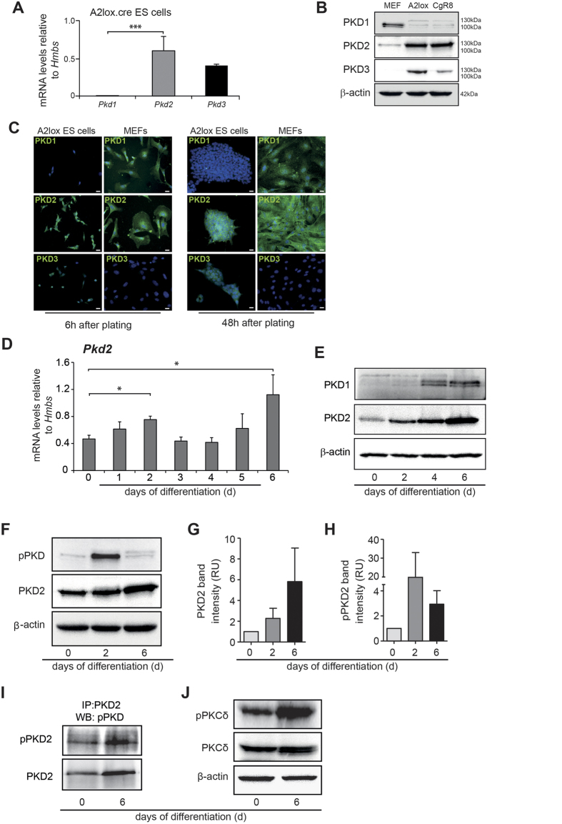
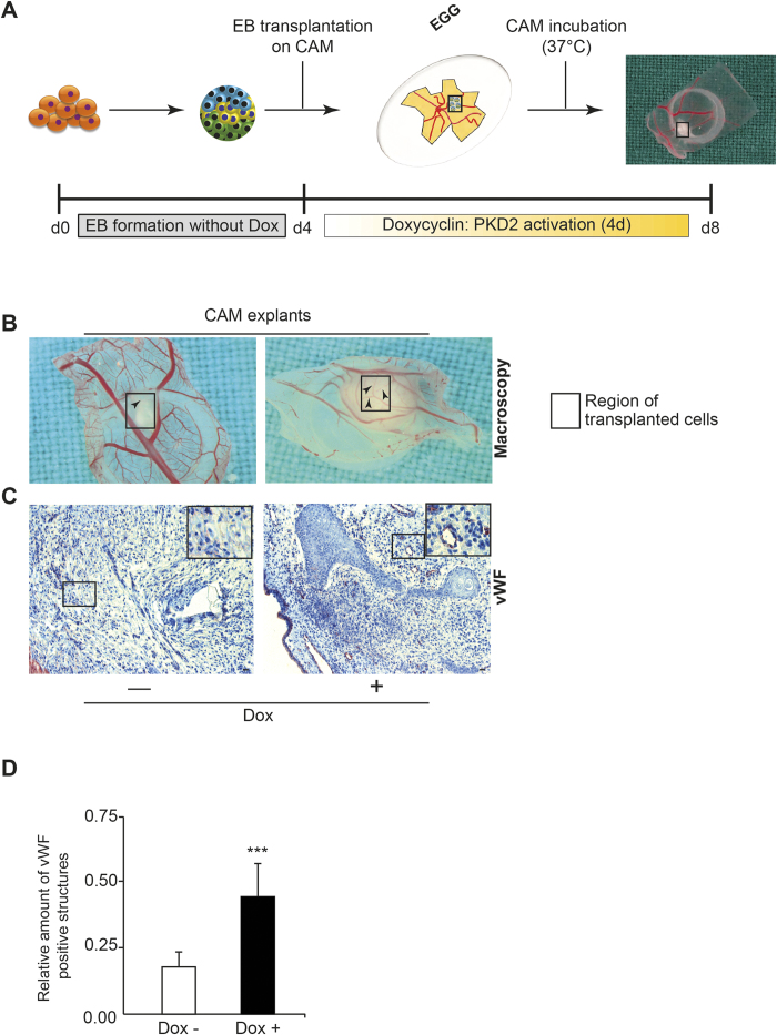
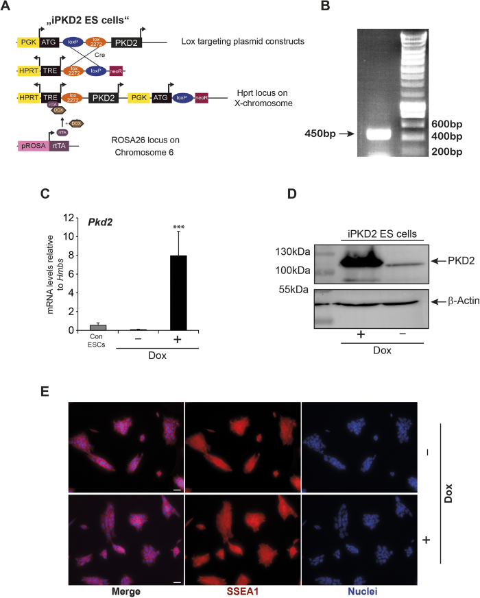
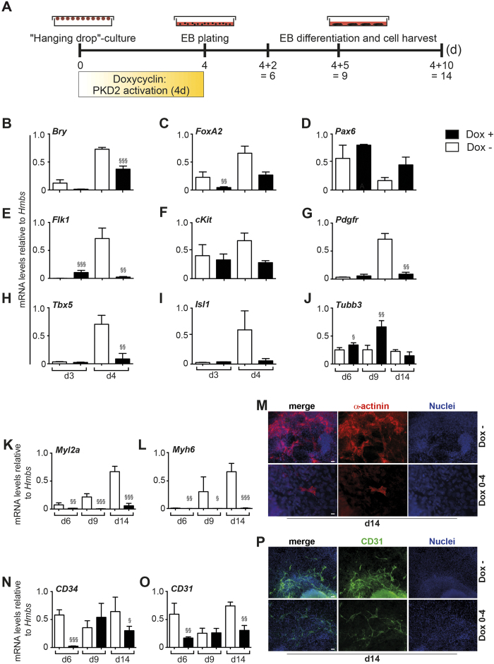
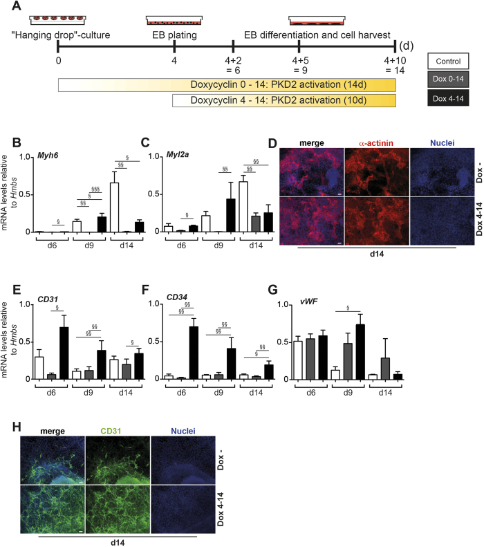
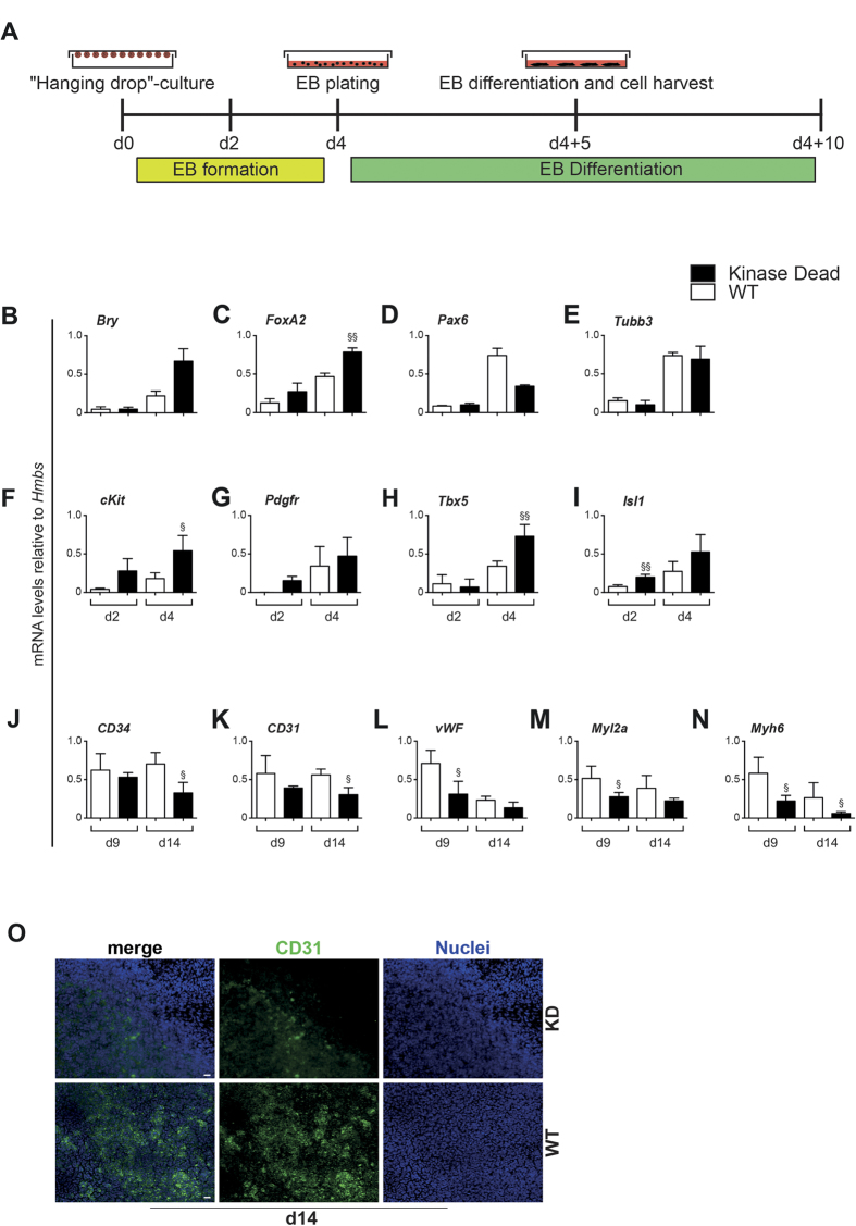
The protein kinase D isoenzymes PKD1/2/3 are prominent downstream targets of PKCs (Protein Kinase Cs) and phospholipase D in various biological systems. Recently, we identified PKD isoforms as novel mediators of tumour cell-endothelial cell communication, tumour cell motility and metastasis. Although PKD isoforms have been implicated in physiological/tumour angiogenesis, a role of PKDs during embryonic development, vasculogenesis and angiogenesis still remains elusive. We investigated the role of PKDs in germ layer segregation and subsequent vasculogenesis and angiogenesis using mouse embryonic stem cells (ESCs). We show that mouse ESCs predominantly express PKD2 followed by PKD3 while PKD1 displays negligible levels. Furthermore, we demonstrate that PKD2 is specifically phosphorylated/activated at the time of germ layer segregation. Time-restricted PKD2-activation limits mesendoderm formation and subsequent cardiovasculogenesis during early differentiation while leading to branching angiogenesis during late differentiation. In line, PKD2 loss-of-function analyses showed induction of mesendodermal differentiation in expense of the neuroectodermal germ layer. Our in vivo findings demonstrate that embryoid bodies transplanted on chicken chorioallantoic membrane induced an angiogenic response indicating that timed overexpression of PKD2 from day 4 onwards leads to augmented angiogenesis in differentiating ESCs. Taken together, our results describe novel and time-dependent facets of PKD2 during early cell fate determination.
蛋白激酶D同工酶PKD1/2/3是蛋白激酶C(PKCs)和磷脂酶D在各种生物系统中的主要下游靶点。最近,我们将PKD同工型鉴定为肿瘤细胞-内皮细胞通讯、肿瘤细胞运动和转移的新型介质。尽管PKD同工型与生理/肿瘤血管生成有关,但PKD在胚胎发育、血管发生和血管生成过程中的作用仍不清楚。我们使用小鼠胚胎干细胞(ESC)研究了PKD在胚层分离以及随后的血管发生和血管生成中的作用。我们发现,小鼠ESC主要表达PKD2,其次是PKD3,而PKD1的表达水平可忽略不计。此外,我们证明PKD2在胚层分离时被特异性磷酸化/激活。限时激活PKD2会限制早期分化过程中中胚层的形成以及随后的心血管发生,同时在晚期分化过程中导致分支血管生成。同样,PKD2功能丧失分析表明,以神经外胚层胚层为代价诱导了中胚层分化。我们的体内研究结果表明,移植到鸡绒毛尿囊膜上的胚状体诱导了血管生成反应,这表明从第4天开始定时过表达PKD2会导致分化中的ESC血管生成增加。综上所述,我们的结果描述了PKD2在早期细胞命运决定过程中的新的和时间依赖性的方面。