Coudrillier Baptiste, Pijanka Jacek K, Jefferys Joan L, Goel Adhiraj, Quigley Harry A, Boote Craig, Nguyen Thao D
Department of Biomedical Engineering, Georgia Institute of Technology, Atlanta, GA, United States of America.
Structural Biophysics Group, School of Optometry and Vision Sciences, Cardiff University, Cardiff, United Kingdom.
PLoS One. 2015 Jul 10;10(7):e0131396. doi: 10.1371/journal.pone.0131396. eCollection 2015.
The biomechanical behavior of the sclera determines the level of mechanical insult from intraocular pressure to the axons and tissues of the optic nerve head, as is of interest in glaucoma. In this study, we measure the collagen fiber structure and the strain response, and estimate the material properties of glaucomatous and normal human donor scleras.
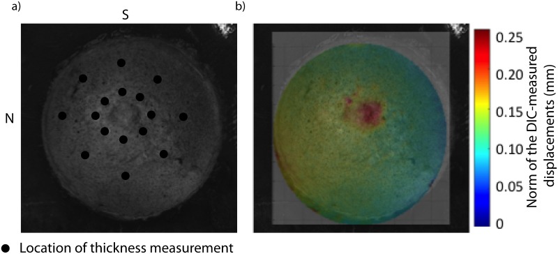
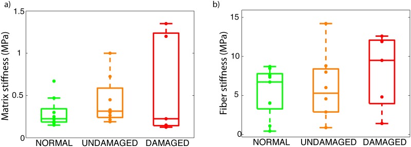
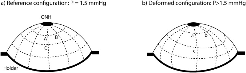
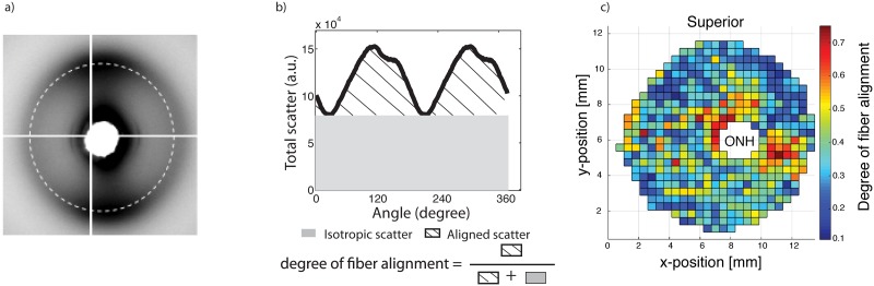
Twenty-two posterior scleras from normal and diagnosed glaucoma donors were obtained from an eyebank. Optic nerve cross-sections were graded to determine the presence of axon loss. The specimens were subjected to pressure-controlled inflation testing. Full-field displacement maps were measured by digital image correlation (DIC) and spatially differentiated to compute surface strains. Maps of the collagen fiber structure across the posterior sclera of each inflated specimen were obtained using synchrotron wide-angle X-ray scattering (WAXS). Finite element (FE) models of the posterior scleras, incorporating a specimen-specific representation of the collagen structure, were constructed from the DIC-measured geometry. An inverse finite element analysis was developed to estimate the stiffness of the collagen fiber and inter-fiber matrix.
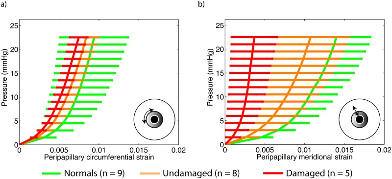
The differences between glaucoma and non-glaucoma eyes were small in magnitude. Sectorial variations of degree of fiber alignment and peripapillary scleral strain significantly differed between normal and diagnosed glaucoma specimens. Meridional strains were on average larger in diagnosed glaucoma eyes compared with normal specimens. Non-glaucoma specimens had on average the lowest matrix and fiber stiffness, followed by undamaged glaucoma eyes, and damaged glaucoma eyes but the differences in stiffness were not significant.
The observed biomechanical and microstructural changes could be the result of tissue remodeling occuring in glaucoma and are likely to alter the mechanical environment of the optic nerve head and contribute to axonal damage.
巩膜的生物力学行为决定了眼内压对视神经乳头轴突和组织的机械损伤程度,这在青光眼研究中备受关注。在本研究中,我们测量了胶原纤维结构和应变响应,并估计了青光眼患者和正常人类供体巩膜的材料特性。
从眼库获取22个正常和已诊断为青光眼的供体的后部巩膜。对视神经横截面进行分级以确定轴突损失的存在。对标本进行压力控制的充气测试。通过数字图像相关(DIC)测量全场位移图,并进行空间微分以计算表面应变。使用同步加速器广角X射线散射(WAXS)获得每个充气标本后部巩膜的胶原纤维结构图谱。根据DIC测量的几何形状构建后部巩膜的有限元(FE)模型,其中包含胶原结构的标本特异性表示。开展逆有限元分析以估计胶原纤维和纤维间基质的刚度。
青光眼和非青光眼眼睛之间的差异幅度较小。正常和已诊断为青光眼的标本之间,纤维排列程度和视乳头周围巩膜应变的扇形变化存在显著差异。与正常标本相比,已诊断为青光眼的眼睛的子午线应变平均更大。非青光眼标本的基质和纤维刚度平均最低,其次是未受损的青光眼眼睛和受损的青光眼眼睛,但刚度差异不显著。
观察到的生物力学和微观结构变化可能是青光眼组织重塑的结果,并且可能会改变视神经乳头的机械环境并导致轴突损伤。