Spain Aisling, Howarth Clare, Khrapitchev Alexandre A, Sharp Trevor, Sibson Nicola R, Martin Chris
Department of Psychology, University of Sheffield, Western Bank, Sheffield S10 2TP, UK; Cancer Research UK & Medical Research Council Oxford Institute for Radiation Oncology, Department of Oncology, University of Oxford, Oxford OX3 7DQ, UK.
Cancer Research UK & Medical Research Council Oxford Institute for Radiation Oncology, Department of Oncology, University of Oxford, Oxford OX3 7DQ, UK.
Neuropharmacology. 2015 Dec;99:210-20. doi: 10.1016/j.neuropharm.2015.07.018. Epub 2015 Jul 17.
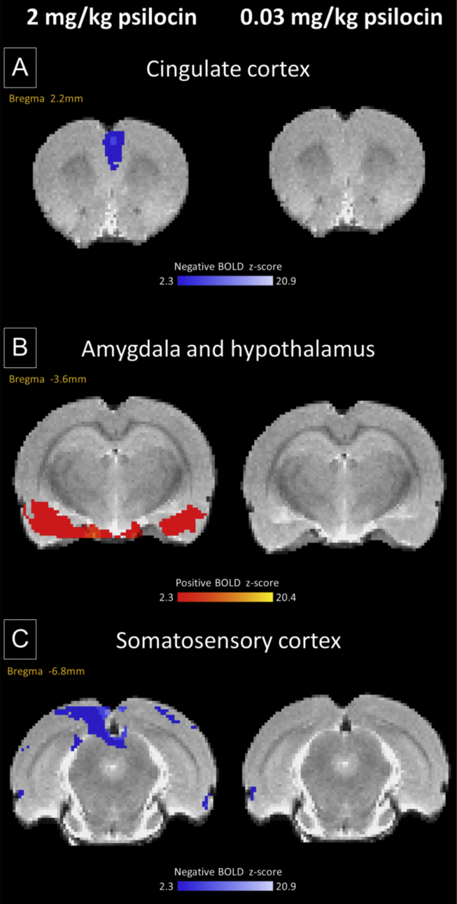
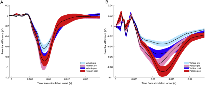
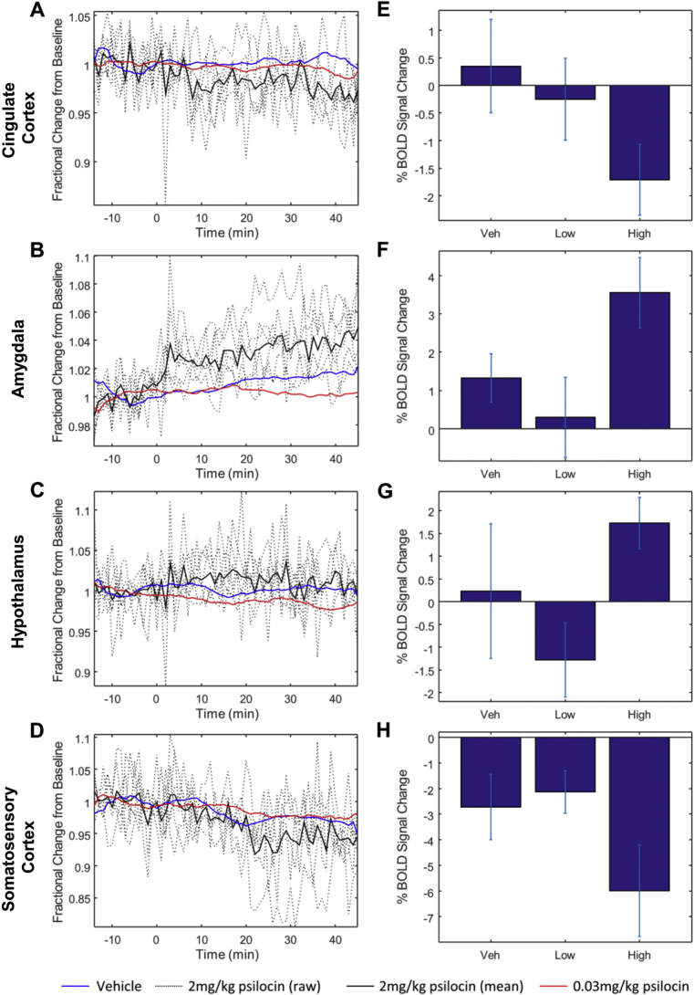
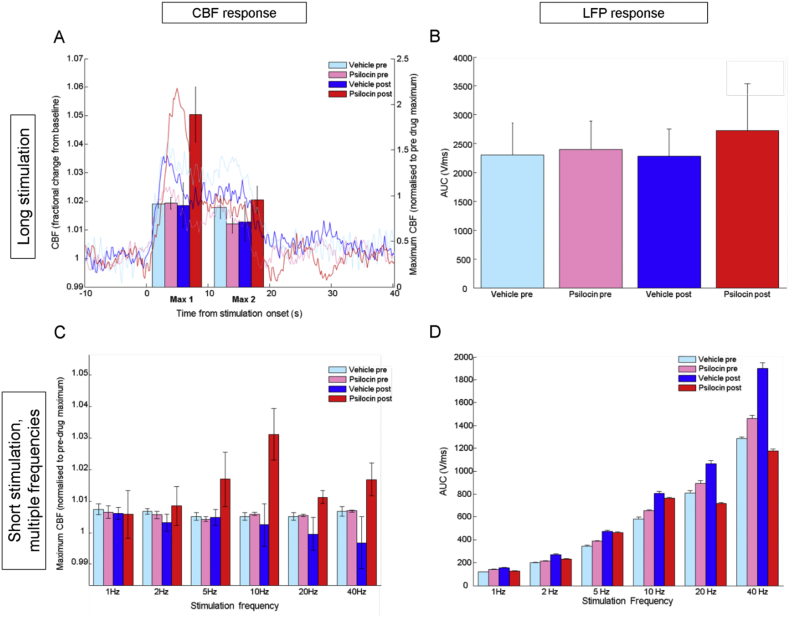
The development of pharmacological magnetic resonance imaging (phMRI) has presented the opportunity for investigation of the neurophysiological effects of drugs in vivo. Psilocin, a hallucinogen metabolised from psilocybin, was recently reported to evoke brain region-specific, phMRI signal changes in humans. The present study investigated the effects of psilocin in a rat model using phMRI and then probed the relationship between neuronal and haemodynamic responses using a multimodal measurement preparation. Psilocin (2 mg/kg or 0.03 mg/kg i.v.) or vehicle was administered to rats (N=6/group) during either phMRI scanning or concurrent imaging of cortical blood flow and recording of local field potentials. Compared to vehicle controls psilocin (2 mg/kg) evoked phMRI signal increases in a number of regions including olfactory and limbic areas and elements of the visual system. PhMRI signal decreases were seen in other regions including somatosensory and motor cortices. Investigation of neurovascular coupling revealed that whilst neuronal responses (local field potentials) to sensory stimuli were decreased in amplitude by psilocin administration, concurrently measured haemodynamic responses (cerebral blood flow) were enhanced. The present findings show that psilocin evoked region-specific changes in phMRI signals in the rat, confirming recent human data. However, the results also suggest that the haemodynamic signal changes underlying phMRI responses reflect changes in both neuronal activity and neurovascular coupling. This highlights the importance of understanding the neurovascular effects of pharmacological manipulations for interpreting haemodynamic neuroimaging data.
药理磁共振成像(phMRI)的发展为体内研究药物的神经生理效应提供了契机。裸盖菇素是从赛洛西宾代谢而来的一种致幻剂,最近有报道称其能在人体中引发脑区特异性的phMRI信号变化。本研究使用phMRI在大鼠模型中研究了裸盖菇素的作用,然后使用多模态测量制剂探究了神经元反应与血流动力学反应之间的关系。在phMRI扫描期间或同时进行皮质血流成像和局部场电位记录时,给大鼠(每组N = 6只)静脉注射裸盖菇素(2 mg/kg或0.03 mg/kg)或赋形剂。与赋形剂对照组相比,裸盖菇素(2 mg/kg)在包括嗅觉和边缘区域以及视觉系统部分区域在内的多个区域引发了phMRI信号增加。在包括体感和运动皮层在内的其他区域观察到phMRI信号降低。对神经血管耦合的研究表明,虽然裸盖菇素给药使对感觉刺激的神经元反应(局部场电位)幅度降低,但同时测量的血流动力学反应(脑血流量)增强。本研究结果表明,裸盖菇素在大鼠中引发了phMRI信号的区域特异性变化,证实了最近的人体数据。然而,结果还表明,phMRI反应背后的血流动力学信号变化反映了神经元活动和神经血管耦合的变化。这突出了理解药物操作的神经血管效应对于解释血流动力学神经成像数据的重要性。