Quak Elske, Le Roux Pierre-Yves, Hofman Michael S, Robin Philippe, Bourhis David, Callahan Jason, Binns David, Desmonts Cédric, Salaun Pierre-Yves, Hicks Rodney J, Aide Nicolas
Nuclear Medicine Department, François Baclesse Cancer Centre, Caen, France.
Nuclear Medicine Department, University Hospital and EA3878 (GETBO) IFR 148, Brest, France.
Eur J Nucl Med Mol Imaging. 2015 Dec;42(13):2072-82. doi: 10.1007/s00259-015-3128-0. Epub 2015 Jul 30.
Point-spread function (PSF) or PSF + time-of-flight (TOF) reconstruction may improve lesion detection in oncologic PET, but can alter quantitation resulting in variable standardized uptake values (SUVs) between different PET systems. This study aims to validate a proprietary software tool (EQ.PET) to harmonize SUVs across different PET systems independent of the reconstruction algorithm used.
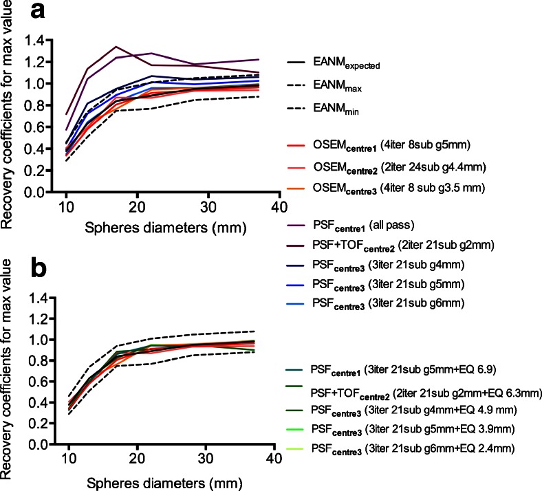
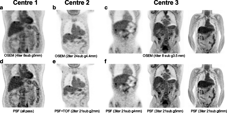
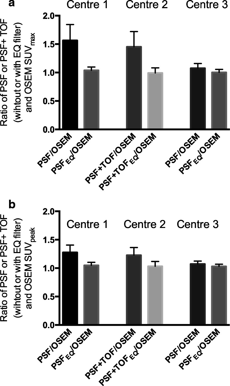
NEMA NU2 phantom data were used to calculate the appropriate filter for each PSF or PSF+TOF reconstruction from three different PET systems, in order to obtain EANM compliant recovery coefficients. PET data from 517 oncology patients were reconstructed with a PSF or PSF+TOF reconstruction for optimal tumour detection and an ordered subset expectation maximization (OSEM3D) reconstruction known to fulfil EANM guidelines. Post-reconstruction, the proprietary filter was applied to the PSF or PSF+TOF data (PSFEQ or PSF+TOFEQ). SUVs for PSF or PSF+TOF and PSFEQ or PSF+TOFEQ were compared to SUVs for the OSEM3D reconstruction. The impact of potential confounders on the EQ.PET methodology including lesion and patient characteristics was studied, as was the adherence to imaging guidelines.
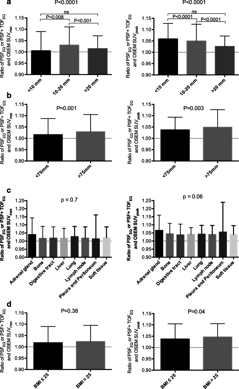
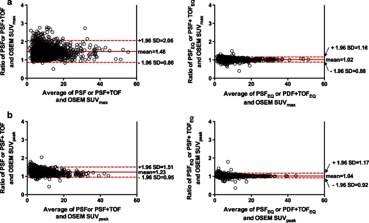
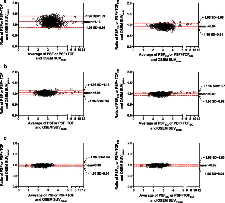
For the 1380 tumour lesions studied, Bland-Altman analysis showed a mean ratio between PSF or PSF+TOF and OSEM3D of 1.46 (95%CI: 0.86-2.06) and 1.23 (95%CI: 0.95-1.51) for SUVmax and SUVpeak, respectively. Application of the proprietary filter improved these ratios to 1.02 (95%CI: 0.88-1.16) and 1.04 (95%CI: 0.92-1.17) for SUVmax and SUVpeak, respectively. The influence of the different confounding factors studied (lesion size, location, radial offset and patient's BMI) was less than 5%. Adherence to the European Association of Nuclear Medicine (EANM) guidelines for tumour imaging was good.
These data indicate that it is not necessary to sacrifice the superior lesion detection and image quality achieved by newer reconstruction techniques in the quest for harmonizing quantitative comparability between PET systems.
点扩散函数(PSF)或PSF+飞行时间(TOF)重建可能会改善肿瘤PET中的病灶检测,但会改变定量结果,导致不同PET系统之间的标准化摄取值(SUV)存在差异。本研究旨在验证一种专有软件工具(EQ.PET),以独立于所使用的重建算法,使不同PET系统的SUV标准化。
使用NEMA NU2体模数据为来自三种不同PET系统的每种PSF或PSF+TOF重建计算合适的滤波器,以获得符合欧洲核医学协会(EANM)标准的恢复系数。对517例肿瘤患者的PET数据进行PSF或PSF+TOF重建以实现最佳肿瘤检测,并进行已知符合EANM指南的有序子集期望最大化(OSEM3D)重建。重建后,将专有滤波器应用于PSF或PSF+TOF数据(PSFEQ或PSF+TOFEQ)。将PSF或PSF+TOF以及PSFEQ或PSF+TOFEQ的SUV与OSEM3D重建的SUV进行比较。研究了包括病灶和患者特征在内的潜在混杂因素对EQ.PET方法的影响,以及对成像指南的遵守情况。
对于所研究的1380个肿瘤病灶,Bland-Altman分析显示,PSF或PSF+TOF与OSEM3D之间SUVmax和SUVpeak的平均比值分别为1.46(95%CI:0.86-2.06)和1.23(95%CI:0.95-1.51)。应用专有滤波器后,SUVmax和SUVpeak的这些比值分别提高到1.02(95%CI:0.88-1.16)和1.04(95%CI:0.92-1.17)。所研究的不同混杂因素(病灶大小、位置、径向偏移和患者体重指数)的影响小于5%。对欧洲核医学协会(EANM)肿瘤成像指南的遵守情况良好。
这些数据表明,在寻求使PET系统之间的定量可比性标准化时,没有必要牺牲更新的重建技术所实现的卓越病灶检测和图像质量。