Bulisani Luís Eduardo Pedigoni, Bulisani Erickson
Brazilian Society of Knee Surgery (SBCJ), São Paulo, SP, Brazil ; Unimed Jundiaí, Jundiaí, SP, Brazil.
Rev Bras Ortop. 2014 Oct 18;49(6):671-4. doi: 10.1016/j.rboe.2014.10.002. eCollection 2014 Nov-Dec.
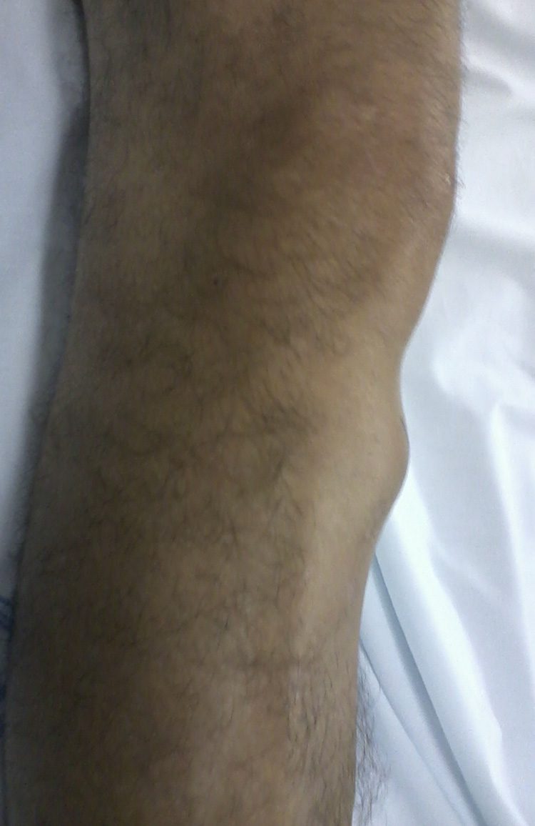
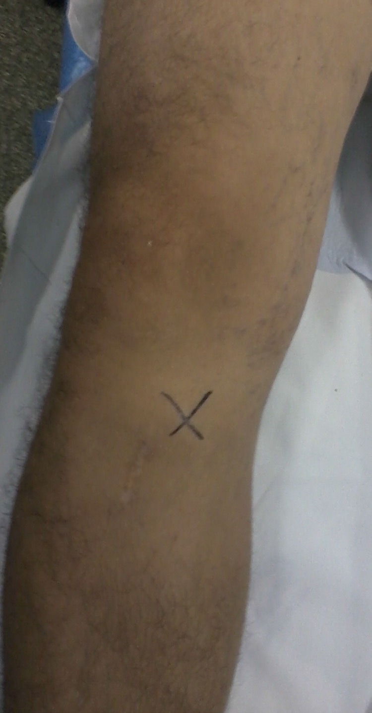
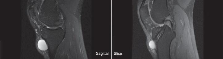
Arthroscopic reconstruction of the anterior cruciate ligament has been modernized through new surgical techniques and new materials. When tibial fixation is performed using an absorbable screw, complications may occur, such as formation of a pre-tibial cyst. The case described here is about a patient who presented an anteromedial synovial cyst in his right knee, three years after having undergone ACL reconstruction. The patient did not present any pain nor any complaints other than a mass that progressively increased in size, worsened after physical activities. Imaging examinations were requested: simple radiography of the knee and magnetic resonance. Anteromedial imaging of the knee showed a mass with well-delimited borders and internal fluid content, suggestive of a synovial cyst, with communication with the joint cavity through the tibial tunnel, without presenting enlargement or absorption of the bone tunnel. The cyst was surgically resected and the tibial tunnel occlusion was performed using a bone plug. The diagnosis of a synovial cyst was subsequently confirmed through the results from the anatomopathological examination. The patient presented good clinical evolution, with disappearance of the symptoms and a return to physical activities.
前交叉韧带的关节镜重建已通过新的手术技术和新材料实现了现代化。当使用可吸收螺钉进行胫骨固定时,可能会出现并发症,如胫骨前囊肿的形成。这里描述的病例是一名患者,在接受前交叉韧带重建三年后,右膝出现了一个滑膜囊肿。除了一个大小逐渐增大、在体育活动后恶化的肿块外,患者没有任何疼痛或不适。进行了影像学检查:膝关节单纯X线摄影和磁共振成像。膝关节的前内侧成像显示一个边界清晰、内部有液体成分的肿块,提示为滑膜囊肿,通过胫骨隧道与关节腔相通,骨隧道未出现扩大或吸收。囊肿通过手术切除,并用骨栓进行胫骨隧道封堵。随后通过解剖病理学检查结果证实了滑膜囊肿的诊断。患者临床进展良好,症状消失并恢复了体育活动。