Chen Wei-Lin, Sheu Joen-Rong, Chen Ray-Jade, Hsiao Shih-Hsin, Hsiao Che-Jen, Chou Yung-Chen, Chung Chi-Li, Hsiao George
Graduate Institute of Medical Sciences and Department of Pharmacology, College of Medicine, Taipei Medical University, Taipei, Taiwan.
Department of Surgery, School of Medicine, College of Medicine, Taipei Medical University, Taipei, Taiwan; Division of General Surgery, Department of Surgery, Taipei Medical University Hospital, Taipei, Taiwan.
PLoS One. 2015 Sep 14;10(9):e0137979. doi: 10.1371/journal.pone.0137979. eCollection 2015.
Tumor necrosis factor (TNF)-α and matrix metalloproteinases (MMPs) are elevated in pleural fluids of tuberculous pleuritis (TBP) where pleural mesothelial cells (PMCs) conduct the first-line defense against Mycobacterium tuberculosis (MTB). However, the clinical implication of TNF-α and MMPs in TBP and the response of PMCs to MTB infection remain unclear.
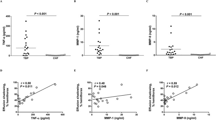
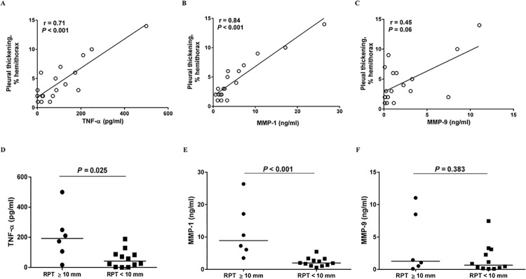
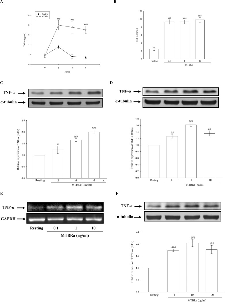
We measured pleural fluid levels of TNF-α and MMPs in patients with TBP (n = 18) or heart failure (n = 18) as controls. Radiological scores for initial effusion amount and residual pleural fibrosis at 6-month follow-up were assessed. In vitro human PMC experiments were performed to assess the effect of heat-killed M. tuberculosis H37Ra (MTBRa) on the expression of TNF-α and MMPs.
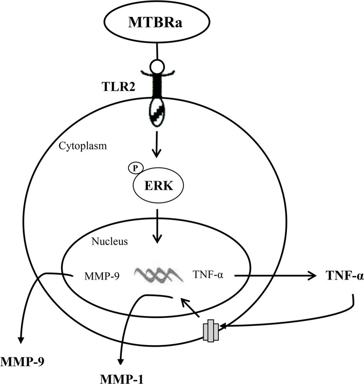
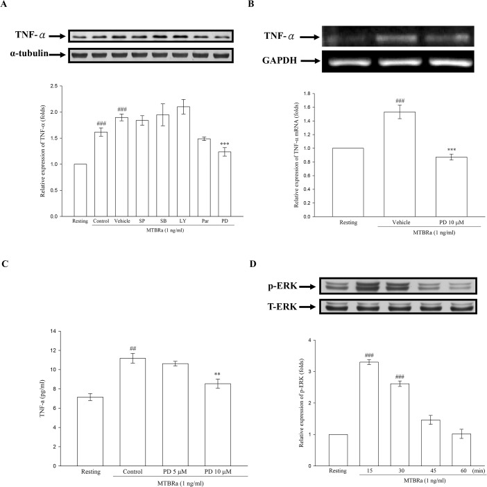
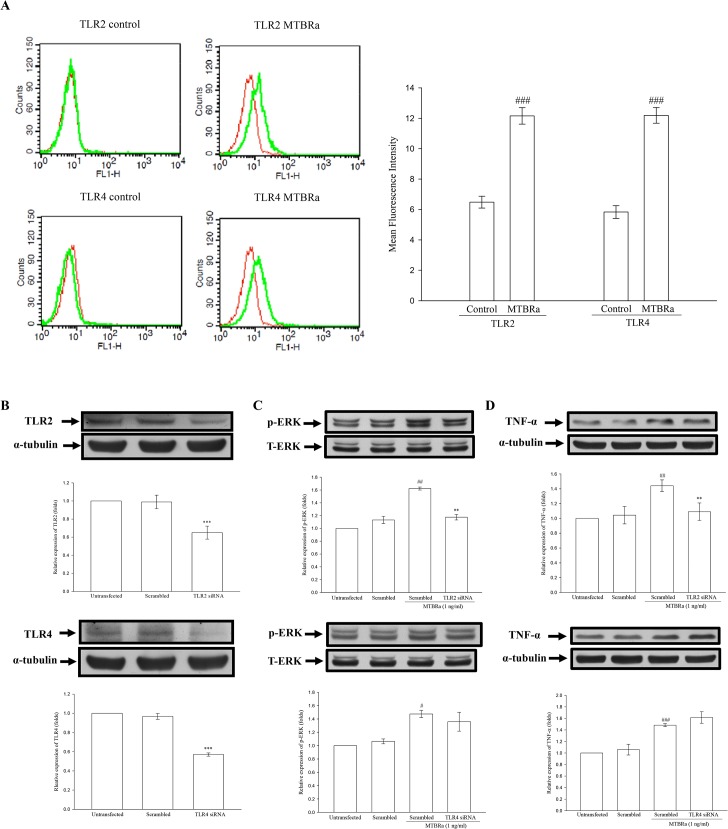
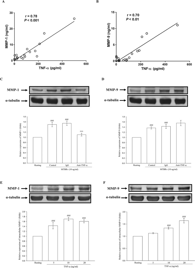
As compared with controls, the effusion levels of TNF-α, MMP-1 and MMP-9 were significantly higher and correlated positively with initial effusion amount in patients with TBP, while TNF-α and MMP-1, but not MMP-9, were positively associated with residual pleural fibrosis of TBP. Moreover, effusion levels of TNF-α had positive correlation with those of MMP-1 and MMP-9 in TBP. In cultured PMCs, MTBRa enhanced TLR2 and TLR4 expression, activated ERK signaling, and upregulated TNF-α mRNA and protein expression. Furthermore, knockdown of TLR2, but not TLR4, significantly inhibited ERK phosphorylation and TNF-α expression. Additionally, both MTBRa and TNF-α markedly induced MMP-1 and MMP-9 synthesis in human PMCs, and TNF-α neutralization substantially reduced the production of MMP-1, but not MMP-9, in response to MTBRa stimulation.
MTBRa activates TLR2/ERK signalings to induce TNF-α and elicit MMP-1 and MMP-9 in human PMCs, which are associated with effusion volume and pleural fibrosis and may contribute to pathogenesis of TBP. Further investigation of manipulation of TNF-α and MMP expression in pleural mesothelium may provide new insights into the mechanisms and rational treatment strategies for TBP.
肿瘤坏死因子(TNF)-α和基质金属蛋白酶(MMPs)在结核性胸膜炎(TBP)患者的胸液中升高,其中胸膜间皮细胞(PMCs)对结核分枝杆菌(MTB)进行一线防御。然而,TNF-α和MMPs在TBP中的临床意义以及PMCs对MTB感染的反应仍不清楚。
我们测量了TBP患者(n = 18)或作为对照的心力衰竭患者(n = 18)胸液中TNF-α和MMPs的水平。评估了初始积液量的放射学评分以及6个月随访时残留的胸膜纤维化情况。进行了体外人PMC实验,以评估热灭活的结核分枝杆菌H37Ra(MTBRa)对TNF-α和MMPs表达的影响。
与对照组相比,TBP患者胸液中TNF-α、MMP-1和MMP-9的水平显著更高,且与初始积液量呈正相关,而TNF-α和MMP-1(而非MMP-9)与TBP的残留胸膜纤维化呈正相关。此外,TBP患者胸液中TNF-α的水平与MMP-1和MMP-9的水平呈正相关。在培养的PMCs中,MTBRa增强了TLR2和TLR4的表达,激活了ERK信号通路,并上调了TNF-α的mRNA和蛋白表达。此外,敲低TLR2(而非TLR4)显著抑制了ERK磷酸化和TNF-α表达。另外,MTBRa和TNF-α均显著诱导人PMCs中MMP-1和MMP-9的合成,并且TNF-α中和显著降低了MTBRa刺激后MMP-1(而非MMP-9)的产生。
MTBRa激活TLR2/ERK信号通路,在人PMCs中诱导TNF-α并引发MMP-1和MMP-9,这与积液量和胸膜纤维化相关,可能有助于TBP的发病机制。进一步研究调节胸膜间皮中TNF-α和MMP表达可能为TBP的机制和合理治疗策略提供新的见解。