Petrillo Mario, Fusco Roberta, Catalano Orlando, Sansone Mario, Avallone Antonio, Delrio Paolo, Pecori Biagio, Tatangelo Fabiana, Petrillo Antonella
Division of Radiology, Department of Diagnostic Imaging, Radiant and Metabolic Therapy, Istituto Nazionale Tumori-IRCCS "Fondazione Giovanni Pascale", Via Mariano Semmola, 80131 Naples, Italy.
Department of Electrical Engineering and Information Technologies, University of Naples Federico II, Via Claudio 21, 80125 Naples, Italy.
Biomed Res Int. 2015;2015:514740. doi: 10.1155/2015/514740. Epub 2015 Aug 27.
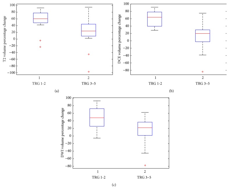
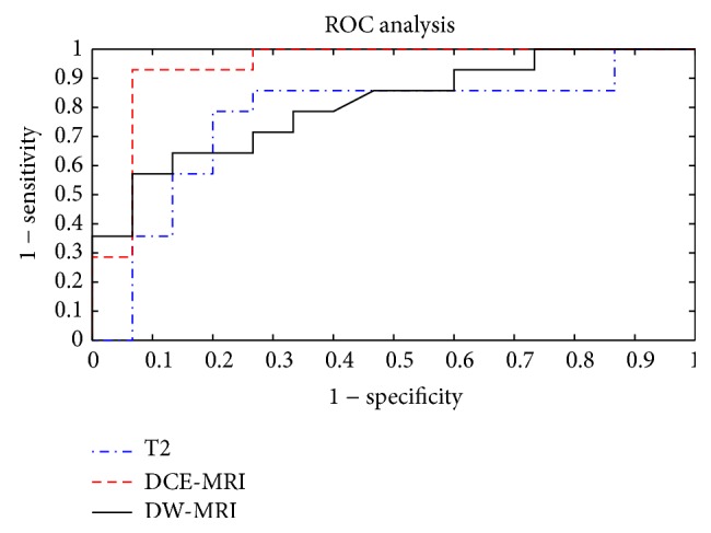
To evaluate MRI for neoadjuvant therapy response assessment in locally advanced rectal cancer (LARC) using dynamic contrast enhanced-MRI (DCE-MRI) and diffusion weighted imaging (DWI), we have compared magnetic resonance volumetry based on DCE-MRI (V(DCE)) and on DWI (V(DWI)) scans with conventional T2-weighted volumetry (V(C)) in LARC patients after neoadjuvant therapy. Twenty-nine patients with LARC underwent MR examination before and after neoadjuvant therapy. A manual segmentation was performed on DCE-MR postcontrast images, on DWI (b-value 800 s/mm(2)), and on conventional T2-weighted images by two radiologists. DCE-MRI, DWI, and T2-weigthed volumetric changes before and after treatment were evaluated. Nonparametric sample tests, interobserver agreement, and receiver operating characteristic curve (ROC) were performed. Diagnostic performance linked to DCE-MRI volumetric change was superior to T2-w and DW-MRI volumetric changes performance (specificity 86%, sensitivity 93%, and accuracy 93%). Area Under ROC (AUC) of V(DCE) was greater than AUCs of V(C) and V(DWI) resulting in an increase of 15.6% and 11.1%, respectively. Interobserver agreement between two radiologists was 0.977, 0.864, and 0.756 for V(C), V(DCE), and V(DWI), respectively. V(DCE) seems to be a promising tool for therapy response assessment in LARC. Further studies on large series of patients are needed to refine technique and evaluate its potential value.
为了使用动态对比增强磁共振成像(DCE-MRI)和扩散加权成像(DWI)评估磁共振成像(MRI)在局部晚期直肠癌(LARC)新辅助治疗反应评估中的应用,我们比较了新辅助治疗后LARC患者基于DCE-MRI(V(DCE))和DWI(V(DWI))扫描的磁共振容积测量与传统T2加权容积测量(V(C))。29例LARC患者在新辅助治疗前后接受了磁共振检查。两名放射科医生对DCE-MR增强后图像、DWI(b值800 s/mm(2))和传统T2加权图像进行了手动分割。评估了治疗前后DCE-MRI、DWI和T2加权容积变化。进行了非参数样本检验、观察者间一致性检验和受试者操作特征曲线(ROC)分析。与DCE-MRI容积变化相关的诊断性能优于T2加权和DW-MRI容积变化性能(特异性86%,敏感性93%,准确性93%)。V(DCE)的ROC曲线下面积(AUC)大于V(C)和V(DWI)的AUC,分别增加了15.6%和11.1%。两名放射科医生对V(C)、V(DCE)和V(DWI)的观察者间一致性分别为0.977、0.864和0.756。V(DCE)似乎是评估LARC治疗反应的一种有前景的工具。需要对大量患者进行进一步研究以完善技术并评估其潜在价值。