• 文献检索
  • 文档翻译
  • 深度研究
  • 学术资讯
  • Suppr Zotero 插件Zotero 插件
  • 邀请有礼
  • 套餐&价格
  • 历史记录
应用&插件
Suppr Zotero 插件Zotero 插件浏览器插件Mac 客户端Windows 客户端微信小程序
定价
高级版会员购买积分包购买API积分包
服务
文献检索文档翻译深度研究API 文档MCP 服务
关于我们
关于 Suppr公司介绍联系我们用户协议隐私条款
关注我们

Suppr 超能文献

核心技术专利:CN118964589B侵权必究
粤ICP备2023148730 号-1Suppr @ 2026

文献检索

告别复杂PubMed语法,用中文像聊天一样搜索,搜遍4000万医学文献。AI智能推荐,让科研检索更轻松。

立即免费搜索

文件翻译

保留排版,准确专业,支持PDF/Word/PPT等文件格式,支持 12+语言互译。

免费翻译文档

深度研究

AI帮你快速写综述,25分钟生成高质量综述,智能提取关键信息,辅助科研写作。

立即免费体验

跗三角骨——纯属偶然还是意义重大?单光子发射计算机断层扫描/计算机断层扫描在一例踝关节撞击病例中的作用

Os Trigonum - Sheer Incidental or Quite Significant? Single Photon Emission Computed Tomography/Computed Tomography's Role in a Case of Ankle Impingement.

作者信息

Chokkappan Kabilan, Srinivasan Sivasubramanian, Subramanian Manickam, Kannivelu Anbalagan

机构信息

Department of Diagnostic Radiology, Khoo Teck Puat Hospital, Singapore.

出版信息

World J Nucl Med. 2015 Sep-Dec;14(3):205-8. doi: 10.4103/1450-1147.163257.

DOI:10.4103/1450-1147.163257
PMID:26420993
原文链接:https://pmc.ncbi.nlm.nih.gov/articles/PMC4564925/
Abstract

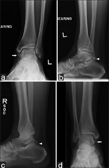
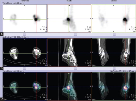
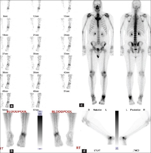
Accessory ossicles are widely prevalent in the ankle and foot. Although they are often asymptomatic, they can present clinically with symptoms at times. When they occur bilaterally in a patient who presents with unilateral complaints, it is clinically difficult to attribute the symptoms to the presence of these common anatomic variants. One needs specific imaging to assess the clinical relevance of the accessory ossicles, in order to tailor the treatment plan. The case presented in this article is one such example, where the patient presented with chronic unilateral ankle pain and initial radiographs revealed bilateral os trigonum and os subtibiale. He underwent a technetium-99m methyl diphosphonate (Tc-99m MDP) bone scan and single photon emission computed tomography/computed tomography (SPECT/CT). The Tc-99m MDP scan showed a focal uptake in the ankle of concern. SPECT/CT complemented the finding by exactly localizing the uptake to the posterior subtalar joint and around the os trigonum, thereby pointing to the diagnosis of os trigonum syndrome.

摘要

副骨在踝关节和足部广泛存在。尽管它们通常无症状,但有时也会出现临床症状。当它们在出现单侧症状的患者中双侧出现时,临床上很难将症状归因于这些常见的解剖变异的存在。为了制定治疗方案,需要特定的影像学检查来评估副骨的临床相关性。本文介绍的病例就是这样一个例子,患者表现为慢性单侧踝关节疼痛,最初的X线片显示双侧距三角骨和胫下骨。他接受了锝-99m亚甲基二膦酸盐(Tc-99m MDP)骨扫描和单光子发射计算机断层扫描/计算机断层扫描(SPECT/CT)。Tc-99m MDP扫描显示踝关节有一处可疑的局部摄取。SPECT/CT通过将摄取精确地定位到距下后关节和距三角骨周围,补充了这一发现,从而指向距三角骨综合征的诊断。

https://cdn.ncbi.nlm.nih.gov/pmc/blobs/153c/4564925/5fed0b4f781b/WJNM-14-205-g003.jpg
https://cdn.ncbi.nlm.nih.gov/pmc/blobs/153c/4564925/5162429906c5/WJNM-14-205-g001.jpg
https://cdn.ncbi.nlm.nih.gov/pmc/blobs/153c/4564925/839ac554a9ef/WJNM-14-205-g002.jpg
https://cdn.ncbi.nlm.nih.gov/pmc/blobs/153c/4564925/5fed0b4f781b/WJNM-14-205-g003.jpg
https://cdn.ncbi.nlm.nih.gov/pmc/blobs/153c/4564925/5162429906c5/WJNM-14-205-g001.jpg
https://cdn.ncbi.nlm.nih.gov/pmc/blobs/153c/4564925/839ac554a9ef/WJNM-14-205-g002.jpg
https://cdn.ncbi.nlm.nih.gov/pmc/blobs/153c/4564925/5fed0b4f781b/WJNM-14-205-g003.jpg

相似文献

1
Os Trigonum - Sheer Incidental or Quite Significant? Single Photon Emission Computed Tomography/Computed Tomography's Role in a Case of Ankle Impingement.跗三角骨——纯属偶然还是意义重大?单光子发射计算机断层扫描/计算机断层扫描在一例踝关节撞击病例中的作用
World J Nucl Med. 2015 Sep-Dec;14(3):205-8. doi: 10.4103/1450-1147.163257.
2
The Utility of Tc-99m Hydroxymethylene Diphosphonate Single-photon Emission Computed Tomography/Computed Tomography in Symptomatic Os Trigonum.锝-99m亚甲基二膦酸盐单光子发射计算机断层扫描/计算机断层扫描在有症状的三角骨中的应用
Indian J Nucl Med. 2018 Apr-Jun;33(2):177-179. doi: 10.4103/ijnm.IJNM_165_17.
3
Prevalence of Os Trigonum on CT Imaging.CT成像上三角骨的患病率。
Foot Ankle Int. 2018 Mar;39(3):338-342. doi: 10.1177/1071100717740937. Epub 2017 Dec 22.
4
The os trigonum syndrome: use of bone scan in the diagnosis.
J Trauma. 1984 Aug;24(8):761-4.
5
Geometrical characteristics of the symptomatic and non-symptomatic ossa trigona. symptomatic 和 non-symptomatic ossa trigona 的几何特征。
Foot Ankle Surg. 2021 Aug;27(6):650-654. doi: 10.1016/j.fas.2020.08.006. Epub 2020 Aug 25.
6
Bone scintigraphic findings of os trigonum: a prospective study of 100 soldiers on active duty.
Foot Ankle Int. 2000 Oct;21(10):822-4. doi: 10.1177/107110070002101005.
7
A Preliminary Study of Ankle Single Photon Emission Computed Tomography/Computed Tomography in Patients With Bony Impingement Syndrome: Association With the Visual Analogue Scale Pain Score.踝关节单光子发射计算机断层扫描/计算机断层扫描在骨撞击综合征患者中的初步研究:与视觉模拟评分疼痛评分的关联
J Foot Ankle Surg. 2019 May;58(3):434-440. doi: 10.1053/j.jfas.2018.09.010. Epub 2019 Mar 13.
8
Potential role of multislice SPECT/CT in impingement syndrome and soft-tissue pathology of the ankle and foot.多层SPECT/CT在踝关节和足部撞击综合征及软组织病变中的潜在作用。
Nucl Med Commun. 2013 Feb;34(2):130-9. doi: 10.1097/MNM.0b013e32835c0964.
9
Accessory ossicles of the foot-an imaging conundrum.足部副骨——影像学难题
Emerg Radiol. 2019 Aug;26(4):465-478. doi: 10.1007/s10140-019-01688-x. Epub 2019 Apr 8.
10
Os trigonum: a discussion of its role in posterior ankle impingement syndrome and a meta-analysis of its prevalence.距骨三角骨:关于其在踝关节后方撞击综合征中作用的讨论及患病率的荟萃分析
Surg Radiol Anat. 2024 Jul;46(7):1137-1143. doi: 10.1007/s00276-024-03389-8. Epub 2024 May 23.

引用本文的文献

1
Male National Basketball Association G-League and Collegiate Basketball Athletes Have a High Prevalence of Radiographic Ankle Abnormalities.美国职业篮球联赛(NBA)发展联盟和大学篮球联赛的男性运动员中,踝关节影像学异常的患病率很高。
Arthrosc Sports Med Rehabil. 2024 Jul 26;6(6):100980. doi: 10.1016/j.asmr.2024.100980. eCollection 2024 Dec.
2
Delayed diagnosis of posterior ankle impingement in pediatric and adolescent patients: does radiology play a role?儿童和青少年患者的后踝撞击征的延迟诊断:影像学是否发挥作用?
Pediatr Radiol. 2020 Feb;50(2):216-223. doi: 10.1007/s00247-019-04547-6. Epub 2019 Nov 9.
3
Anatomical variation in the ankle and foot: from incidental finding to inductor of pathology. Part I: ankle and hindfoot.

本文引用的文献

1
Posterior ankle impingement in dancers and athletes.舞者和运动员的后踝撞击综合征
Foot Ankle Clin. 2013 Jun;18(2):301-18. doi: 10.1016/j.fcl.2013.02.008.
2
Potential role of multislice SPECT/CT in impingement syndrome and soft-tissue pathology of the ankle and foot.多层SPECT/CT在踝关节和足部撞击综合征及软组织病变中的潜在作用。
Nucl Med Commun. 2013 Feb;34(2):130-9. doi: 10.1097/MNM.0b013e32835c0964.
3
MR imaging of ankle impingement syndromes.踝关节撞击综合征的磁共振成像
踝关节和足部的解剖变异:从偶然发现到病理诱发因素。第一部分:踝关节和后足。
Insights Imaging. 2019 Jul 31;10(1):74. doi: 10.1186/s13244-019-0746-2.
AJR Am J Roentgenol. 2003 Aug;181(2):551-9. doi: 10.2214/ajr.181.2.1810551.
4
Posterior ankle impingement syndrome: MR imaging findings in seven patients.后踝撞击综合征:7例患者的磁共振成像表现
Radiology. 2000 May;215(2):497-503. doi: 10.1148/radiology.215.2.r00ma01497.
5
Lower extremity scintigraphy: the foot and ankle.下肢闪烁扫描术:足部和脚踝
Semin Nucl Med. 1998 Jan;28(1):62-77. doi: 10.1016/s0001-2998(98)80019-6.
6
The os trigonum syndrome: imaging features.距骨三角骨综合征:影像学特征
AJR Am J Roentgenol. 1996 Jan;166(1):125-9. doi: 10.2214/ajr.166.1.8571860.
7
Posterior ankle impingement.
Foot Ankle Int. 1994 Jan;15(1):2-8. doi: 10.1177/107110079401500102.
8
Posterior intermalleolar ligament of the ankle: normal anatomy and MR imaging features.踝关节后踝间韧带:正常解剖及磁共振成像特征
AJR Am J Roentgenol. 1995 Aug;165(2):387-90. doi: 10.2214/ajr.165.2.7618563.
9
The os trigonum syndrome: use of bone scan in the diagnosis.
J Trauma. 1984 Aug;24(8):761-4.