Kohno Hideo, Koso Hideto, Okano Kiichiro, Sundermeier Thomas R, Saito Saburo, Watanabe Sumiko, Tsuneoka Hiroshi, Sakai Tsutomu
Department of Ophthalmology, The Jikei University School of Medicine, 105-8461, Tokyo, Japan.
Tokyu Hospital, 145-0062, Tokyo, Japan.
J Neuroinflammation. 2015 Oct 12;12:188. doi: 10.1186/s12974-015-0408-3.
Though accumulating evidence suggests that microglia, resident macrophages in the retina, and bone marrow-derived macrophages can cause retinal inflammation which accelerates photoreceptor cell death, the details of how these cells are activated during retinal degeneration (RD) remain uncertain. Therefore, it is important to clarify which cells play a dominant role in fueling retinal inflammation. However, distinguishing between microglia and macrophages is difficult using conventional techniques such as cell markers (e.g., Iba-1). Recently, two mouse models for visualizing chemokine receptors were established, Cx3cr1 (GFP/GFP) and Ccr2 (RFP/RFP) mice. As Cx3cr1 is expressed in microglia and Ccr2 is reportedly expressed in activated macrophages, these mice have the potential to distinguish microglia and macrophages, yielding novel information about the activation of these inflammatory cells and their individual roles in retinal inflammation.
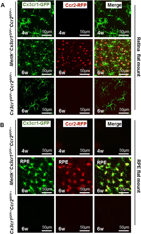
In this study, c-mer proto-oncogene tyrosine kinase (Mertk) (-/-) mice, which show photoreceptor cell death due to defective retinal pigment epithelium phagocytosis, were employed as an animal model of RD. Mertk (-/-) Cx3cr1 (GFP/+) Ccr2 (RFP/+) mice were established by breeding Mertk (-/-) , Cx3cr1 (GFP/GFP) , and Ccr2 (RFP/RFP) mice. The retinal morphology and pattern of inflammatory cell activation and invasion of Mertk (-/-) Cx3cr1 (GFP/+) Ccr2 (RFP/+) mice were evaluated using retina and retinal pigment epithelium (RPE) flat mounts, retinal sections, and flow cytometry.
Four-week-old Mertk (-/-) Cx3cr1 (GFP/+) Ccr2 (RFP/+) mice showed Cx3cr1-GFP-positive microglia in the inner retina. Cx3cr1-GFP and Ccr2-RFP dual positive activated microglia were observed in the outer retina and subretinal space of 6- and 8-week-old animals. Ccr2-RFP single positive bone marrow-derived macrophages were observed to migrate into the retina of Mertk (-/-) Cx3cr1 (GFP/+) Ccr2 (RFP/+) mice. These invading cells were still observed in the subretinal space in 18-week-old animals.
Cx3cr1-GFP-positive microglia and Ccr2-RFP-positive macrophages were distinguishable in the retinas of Mertk (-/-) Cx3cr1 (GFP/+) Ccr2 (RFP/+) mice. In addition, Ccr2 expression in Cx3cr1 positive microglia is a feature of microglial activation in RD. Mertk (-/-) Cx3cr1 (GFP/+) Ccr2 (RFP/+) mice enabled observation of microglial activation over time during RD and may be useful for developing inflammation-targeted treatment strategies for RD in the future.
尽管越来越多的证据表明,小胶质细胞(视网膜中的常驻巨噬细胞)和骨髓来源的巨噬细胞可引发视网膜炎症,加速光感受器细胞死亡,但这些细胞在视网膜变性(RD)过程中如何被激活的细节仍不明确。因此,明确哪些细胞在加剧视网膜炎症中起主导作用很重要。然而,使用细胞标志物(如离子钙接头蛋白1,Iba-1)等传统技术很难区分小胶质细胞和巨噬细胞。最近,建立了两种用于可视化趋化因子受体的小鼠模型,即Cx3cr1(绿色荧光蛋白/绿色荧光蛋白)和Ccr2(红色荧光蛋白/红色荧光蛋白)小鼠。由于Cx3cr1在小胶质细胞中表达,且据报道Ccr2在活化的巨噬细胞中表达,这些小鼠有潜力区分小胶质细胞和巨噬细胞,从而产生有关这些炎症细胞激活及其在视网膜炎症中各自作用的新信息。
在本研究中,由于视网膜色素上皮吞噬功能缺陷而出现光感受器细胞死亡的c-mer原癌基因酪氨酸激酶(Mertk)基因敲除小鼠被用作RD的动物模型。通过将Mertk基因敲除小鼠、Cx3cr1(绿色荧光蛋白/绿色荧光蛋白)小鼠和Ccr2(红色荧光蛋白/红色荧光蛋白)小鼠杂交,建立了Mertk基因敲除Cx3cr1(绿色荧光蛋白/ +)Ccr2(红色荧光蛋白/ +)小鼠。使用视网膜和视网膜色素上皮(RPE)铺片、视网膜切片及流式细胞术评估Mertk基因敲除Cx3cr1(绿色荧光蛋白/ +)Ccr2(红色荧光蛋白/ +)小鼠的视网膜形态、炎症细胞激活模式及浸润情况。
4周龄的Mertk基因敲除Cx3cr1(绿色荧光蛋白/ +)Ccr2(红色荧光蛋白/ +)小鼠在内层视网膜中显示出Cx3cr1-绿色荧光蛋白阳性的小胶质细胞。在6周龄和8周龄动物的外层视网膜和视网膜下间隙中观察到Cx3cr1-绿色荧光蛋白和Ccr2-红色荧光蛋白双阳性的活化小胶质细胞。观察到Ccr2-红色荧光蛋白单阳性的骨髓来源巨噬细胞迁移到Mertk基因敲除Cx3cr1(绿色荧光蛋白/ +)Ccr2(红色荧光蛋白/ +)小鼠的视网膜中。在18周龄动物的视网膜下间隙中仍可观察到这些浸润细胞。
在Mertk基因敲除Cx3cr1(绿色荧光蛋白/ +)Ccr2(红色荧光蛋白/ +)小鼠的视网膜中,Cx3cr1-绿色荧光蛋白阳性的小胶质细胞和Ccr2-红色荧光蛋白阳性的巨噬细胞是可区分的。此外,Cx3cr1阳性小胶质细胞中Ccr2的表达是RD中小胶质细胞激活的一个特征。Mertk基因敲除Cx3cr1(绿色荧光蛋白/ +)Ccr2(红色荧光蛋白/ +)小鼠能够观察到RD过程中小胶质细胞随时间的激活情况,可能有助于未来开发针对RD的炎症靶向治疗策略。