Xiao Hai-Tao, Peng Jiao, Hu Dong-Dong, Lin Cheng-Yuan, Du Bin, Tsang Siu-Wai, Lin Ze-Si, Zhang Xiao-Jun, Lueng Feng-Ping, Han Quan-Bin, Bian Zhao-Xiang
School of Chinese Medicine, Hong Kong Baptist University, Hong Kong, Hong Kong ; School of Pharmacy, Guiyang Medical University, Guiyang, 550004 China.
Department of Surgery, LKS Faculty of Medicine, The University of Hong Kong, Hong Kong, Hong Kong.
Chin Med. 2015 Oct 13;10:29. doi: 10.1186/s13020-015-0061-x. eCollection 2015.
Qing-dai powder (QDP), comprising Indigo naturalis (Qing-dai) and dried alum (Ku-fan), was used in Chinese medicine to treat the conditions associated with mucosal hemorrhage, such as ulcerative colitis (UC). This study aims to investigate the effects and potential mechanism of QDP on dextran sulfate sodium (DSS)-induced acute colitis in mice and to examine the regulatory effects of QDP on macrophages.
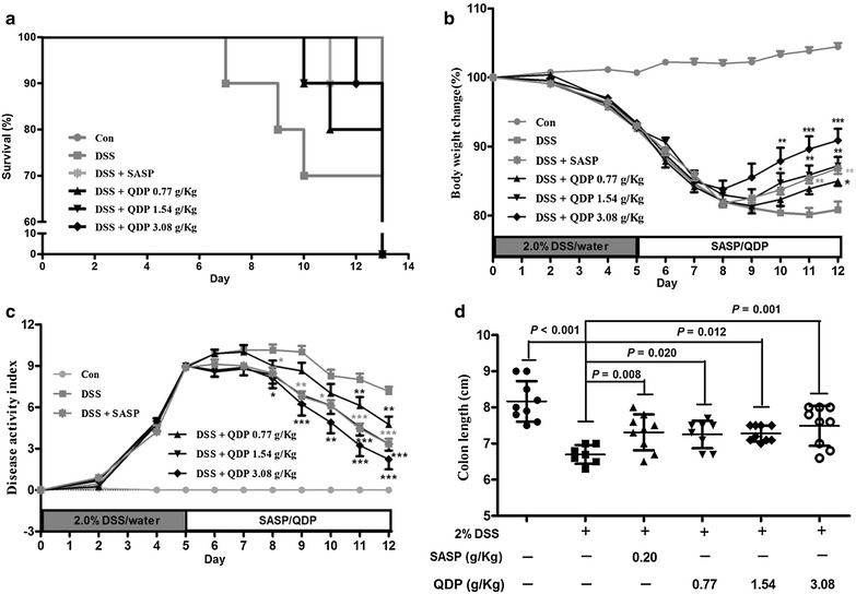
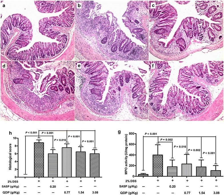
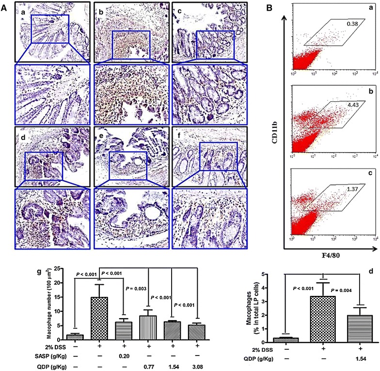
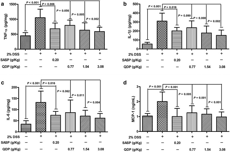
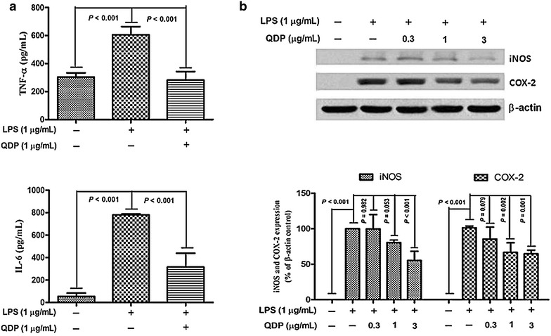
Seven- to eight-week-old male C57BL/6 mice were challenged with 2.0 % DSS in drinking water for 5 days and then the colitic mice were arbitrarily allocated into five groups (n = 10 for each group). QDP (0.77, 1.54 and 3.08 g/kg) and sulfasalazine (SASP) (0.20 g/kg) were orally administered for 7 days. The disease activity index was determined by scores of body weight loss, diarrhea and rectal bleeding; histological signs of damage was analyzed by H&E staining; myeloperoxidase activity was measured by colorimetric method, levels of proinflammatory cytokines were determined by ELISA; changes in macrophages in the colon were analyzed by immunohistochemistry (IHC) and flow cytometry. Lipopolysaccharide (LPS)-induced RAW264.7 cells were treated with or without QDP, then the production of TNF-α and IL-6 were measured by ELISA; and protein molecules such as COX-2, iNOS, IкB-α were determined by Western blot.
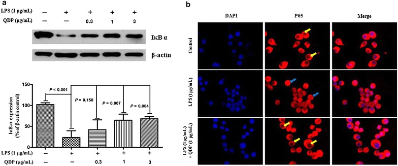
Oral administration of QDP at dosages of 1.54 and 3.08 g/kg significantly reduced disease activity index on day 12 (P < 0.001 for 1.54 g/kg and P < 0.0008 for 3.08 g/kg), colon shortening (P = 0.012 for 1.54 g/kg, P = 0.001 for 3.08 g/kg), histological damage (P < 0.001 for 1.54 g/kg, P < 0.001 for 3.08 g/kg) and colonic myeloperoxidase activity (P = 0.002 for 1.54 g/kg, P < 0.001 for 3.08 g/kg) of DSS-treated mice. Moreover, QDP treatment (1.54 and 3.08 g/kg) significantly decreased DSS-induced infiltration of macrophages, and production of TNF-α (P = 0.005 for 1.54 g/kg, P = 0.002 for 3.08 g/kg), IL-1β (P = 0.008 for 1.54 g/kg, P = 0.002 for 3.08 g/kg) and IL-6 (P = 0.011 for 1.54 g/kg, P = 0.004 for 3.08 g/kg) in colonic tissues, and also reduced serum MCP-1 levels (P = 0.001 for 1.54 g/kg, P < 0.001 for 3.08 g/kg). In RAW264.7 cells, QDP significantly suppressed LPS-induced production of TNF-α and IL-6 (Both P < 0.001 for 1.0 μg/mL QDP treatment) and expression levels of COX-2 (P = 0.002 and P = 0.001 for 1 and 3 μg/mL QDP treatment, respectively) and iNOS (P < 0.001 for 3 μg/mL QDP treatment) by inhibiting IкB-α degradation (P = 0.007 and P = 0.004 for 1 and 3 μg/mL QDP treatment, respectively) and NF-кB p65 nuclear translocation.
QDP suppressed the inflammatory responses of colonic macrophages in DSS-induced UC in mice and LPS-induced RAW264.7 cells.
青黛散(QDP)由青黛(Indigo naturalis)和枯矾(dried alum)组成,在中医中用于治疗与黏膜出血相关的病症,如溃疡性结肠炎(UC)。本研究旨在探讨QDP对葡聚糖硫酸钠(DSS)诱导的小鼠急性结肠炎的影响及潜在机制,并研究QDP对巨噬细胞的调节作用。
将7至8周龄的雄性C57BL/6小鼠用2.0%的DSS饮用水处理5天,然后将患结肠炎的小鼠随机分为五组(每组n = 10)。口服给予QDP(0.77、1.54和3.08 g/kg)和柳氮磺胺吡啶(SASP)(0.20 g/kg),持续7天。通过体重减轻、腹泻和直肠出血评分确定疾病活动指数;通过苏木精-伊红(H&E)染色分析组织损伤的组织学征象;用比色法测量髓过氧化物酶活性;用酶联免疫吸附测定(ELISA)法测定促炎细胞因子水平;通过免疫组织化学(IHC)和流式细胞术分析结肠中巨噬细胞的变化。用或不用QDP处理脂多糖(LPS)诱导的RAW264.7细胞,然后用ELISA法测量TNF-α和IL-6的产生;用蛋白质印迹法测定COX-2、iNOS、IκB-α等蛋白质分子。
口服1.54和3.08 g/kg剂量的QDP可显著降低第12天的疾病活动指数(1.54 g/kg时P < 0.001,3.08 g/kg时P < 0.0008)、结肠缩短(1.54 g/kg时P = 0.012,3.08 g/kg时P = 0.001)、组织学损伤(1.54 g/kg时P < 0.001,3.08 g/kg时P < 0.001)以及DSS处理小鼠的结肠髓过氧化物酶活性(1.54 g/kg时P = 0.002,3.08 g/kg时P < 0.001)。此外,QDP处理(1.54和3.08 g/kg)可显著减少DSS诱导的巨噬细胞浸润,以及结肠组织中TNF-α(1.54 g/kg时P = 0.005,3.08 g/kg时P = 0.002)、IL-1β(1.54 g/kg时P = 0.008,3.08 g/kg时P = 0.002)和IL-6(1.54 g/kg时P = 0.011,3.08 g/kg时P = 0.004)的产生,还可降低血清MCP-1水平(1.54 g/kg时P = 0.001,3.08 g/kg时P < 0.001)。在RAW264.7细胞中,QDP通过抑制IκB-α降解(1和3 μg/mL QDP处理时分别为P = 0.007和P = 0.004)和NF-κB p65核转位,显著抑制LPS诱导的TNF-α和IL-6产生(1.0 μg/mL QDP处理时两者P均< 0.001)以及COX-2(1和3 μg/mL QDP处理时分别为P = 0.002和P = 0.001)和iNOS(3 μg/mL QDP处理时P < 0.001)的表达水平。
QDP可抑制DSS诱导的小鼠UC中结肠巨噬细胞的炎症反应以及LPS诱导的RAW264.7细胞的炎症反应。