Azin Mahdieh, Mirnajafi-Zadeh Javad, Javan Mohammad
Department of Physiology, Faculty of Medical Sciences, Tarbiat Modares University, Tehran, Iran.
Cell J. 2015 Fall;17(3):540-456. doi: 10.22074/cellj.2015.14. Epub 2015 Oct 7.
Hippocampal insults have been observed in multiple sclerosis (MS) patients. Fibroblast growth factor-2 (FGF2) induces neurogenesis in the hippocampus and en- hances the proliferation, migration and differentiation of oligodendrocyte progenitor cells (OPCs). In the current study, we have investigated the effect of FGF2 on the processes of gliotoxin induced demyelination and subsequent remyelination in the hippocampus.
In this experimental study adult male Sprague-Dawley rats re- ceived either saline or lysolecithin (LPC) injections to the right hippocampi. Animals re- ceived intraperitoneal (i.p.) injections of FGF2 (5 ng/g) on days 0, 5, 12 and 26 post-LPC. Expressions of myelin basic protein (Mbp) as a marker of myelination, Olig2 as a marker of OPC proliferation, Nestin as a marker of neural progenitor cells, and glial fibrillary acidic protein (Gfap) as a marker of reactive astrocytes were investigated in the right hippocampi by reverse transcriptase-polymerase chain reaction (RT-PCR).
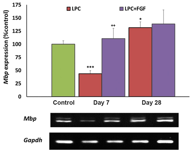
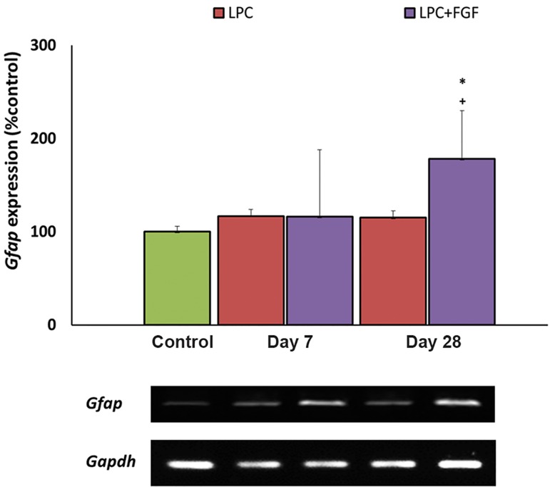
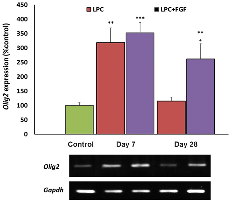
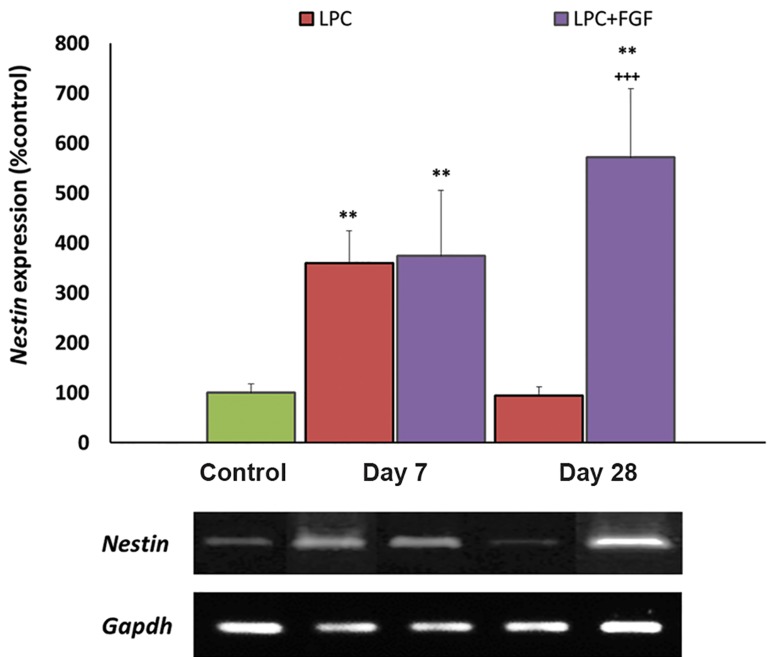
There was reduced Mbp expression at seven days after LPC injection, in- creased expressions of Olig2 and Nestin, and the level of Gfap did not change. FGF2 treatment reversed the expression level of Mbp to the control, significantly enhanced the levels of Olig2 and Nestin, but did not change the level of Gfap. At day-28 post- LPC, the expression level of Mbp was higher than the control in LPC-treated animals that received FGF2. The levels of Olig2, Nestin and Gfap were at the control level in the non-treated LPC group but significantly higher in the FGF2-treated LPC group.
FGF2 enhanced hippocampal myelination and potentiated the recruitment of OPCs and neural stem cells (NSCs) to the lesion area. Long-term application of FGF2 might also enhance astrogliosis in the lesion site.
在多发性硬化症(MS)患者中已观察到海马损伤。成纤维细胞生长因子-2(FGF2)可诱导海马神经发生,并增强少突胶质前体细胞(OPC)的增殖、迁移和分化。在本研究中,我们调查了FGF2对海马中由神经毒素诱导的脱髓鞘及随后的髓鞘再生过程的影响。
在本实验研究中,成年雄性Sprague-Dawley大鼠右侧海马接受生理盐水或溶血卵磷脂(LPC)注射。在LPC注射后的第0、5、12和26天,动物接受腹腔内(i.p.)注射FGF2(5 ng/g)。通过逆转录聚合酶链反应(RT-PCR)研究右侧海马中作为髓鞘形成标志物的髓鞘碱性蛋白(Mbp)、作为OPC增殖标志物的Olig2、作为神经祖细胞标志物的巢蛋白(Nestin)以及作为反应性星形胶质细胞标志物的胶质纤维酸性蛋白(Gfap)的表达。
LPC注射后7天,Mbp表达降低,Olig2和Nestin表达增加,Gfap水平未改变。FGF2治疗使Mbp表达水平恢复至对照水平,显著提高了Olig2和Nestin水平,但未改变Gfap水平。在LPC注射后第28天,接受FGF2治疗的LPC处理动物中Mbp表达水平高于对照。在未处理的LPC组中,Olig2、Nestin和Gfap水平处于对照水平,但在FGF2处理的LPC组中显著更高。
FGF2增强了海马髓鞘形成,并促进了OPC和神经干细胞(NSC)向损伤区域的募集。长期应用FGF2可能还会增强损伤部位的星形胶质细胞增生。