Saarinen Irena, Mirtti Tuomas, Seikkula Heikki, Boström Peter J, Taimen Pekka
Department of Pathology, University of Turku and Turku University Hospital, Turku, Finland; MediCity, Research Laboratory, University of Turku, Turku, Finland.
Department of Pathology, Helsinki University Hospital and Finnish Institute for Molecular Medicine, University of Helsinki, Helsinki, Finland.
PLoS One. 2015 Oct 15;10(10):e0140671. doi: 10.1371/journal.pone.0140671. eCollection 2015.
Prostate cancer (PCa) is the most common cancer among men in western countries. While active surveillance is increasingly utilized, the majority of patients are currently treated with radical prostatectomy. In order to avoid over-treatment, there is an indisputable need for reliable biomarkers to identify the potentially aggressive and lethal cases. Nuclear intermediate filament proteins called lamins play a role in chromatin organization, gene expression and cell stiffness. The expression of lamin A is associated with poor outcome in colorectal cancer but to date the prognostic value of the lamins has not been tested in other solid tumors.
We studied the expression of different lamins with immunohistochemistry in a tissue microarray material of 501 PCa patients undergoing radical prostatectomy and lymph node dissection. Patients were divided into two staining categories (low and high expression). The correlation of lamin expression with clinicopathological variables was tested and the association of lamin status with biochemical recurrence (BCR) and disease specific survival (DSS) was further analyzed.
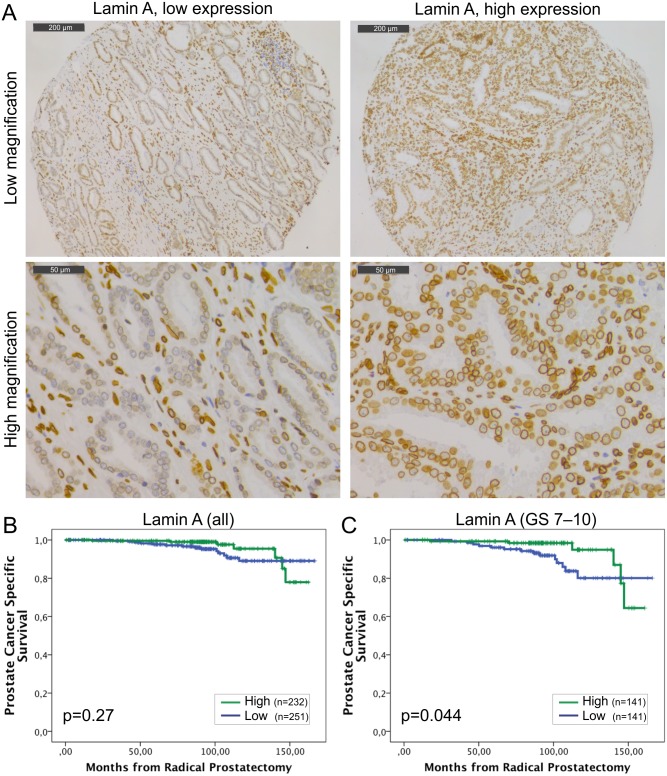
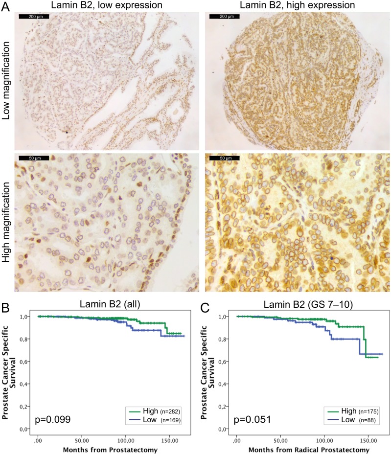
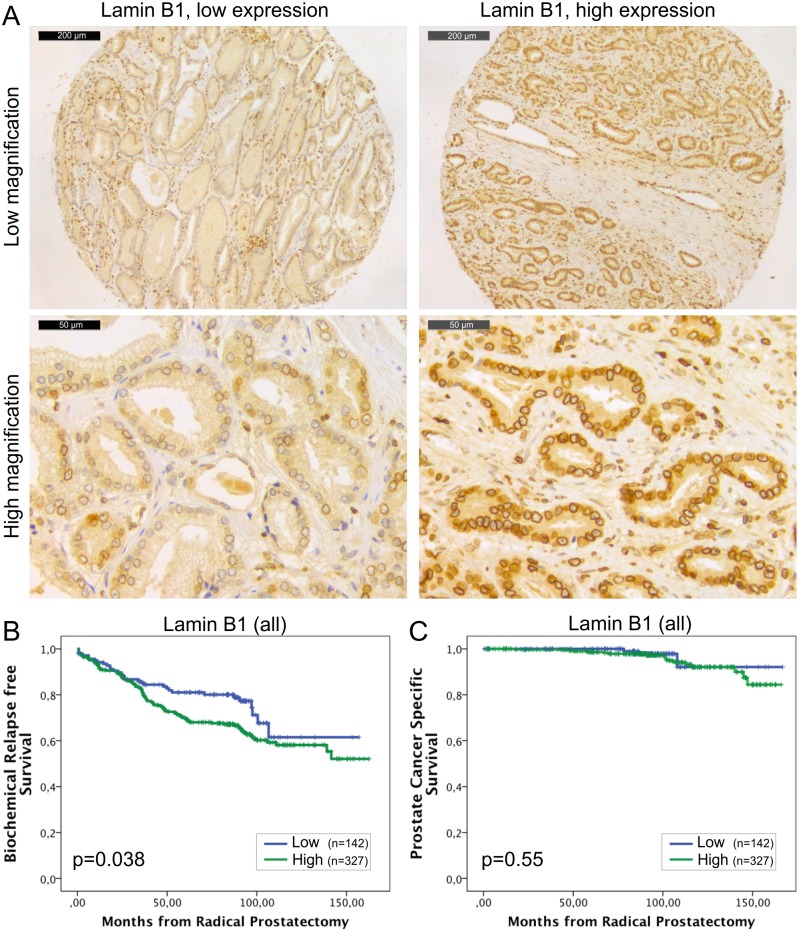
Low expression of lamin A associated with lymph node positivity (p<0.01) but not with other clinicopathological variables and low expression had a borderline independent significant association with DSS (HR = 0.4; 95% CI 0.2-1.0; p = 0.052). Similarly, low lamin C expression associated with poorer survival (HR = 0.2; 95% CI 0.1-0.6; p = 0.004). Lamin B1 expression did not associate with clinicopathological variables but high expression independently predicted BCR in multivariable Cox regression analysis (HR = 1.8; 95% CI 1.1-2.9; p = 0.023). Low expression of lamin B2 correlated with lymph node positivity (p<0.01) and predicted unfavorable DSS (HR = 0.4; 95% CI 0.2-1.0; p = 0.047).
These results suggest differential roles for lamins in PCa progression. Reduced amounts of lamin A/C and B2 increase risk for lymph node metastasis and disease specific death possibly through increased nuclear deformability while high expression of lamin B1 predicts disease recurrence.
前列腺癌(PCa)是西方国家男性中最常见的癌症。虽然主动监测的应用越来越广泛,但目前大多数患者仍接受根治性前列腺切除术治疗。为了避免过度治疗,显然需要可靠的生物标志物来识别潜在侵袭性和致命性病例。称为核纤层蛋白的核中间丝蛋白在染色质组织、基因表达和细胞硬度方面发挥作用。核纤层蛋白A的表达与结直肠癌的不良预后相关,但迄今为止,核纤层蛋白在其他实体瘤中的预后价值尚未得到检验。
我们采用免疫组织化学方法研究了501例接受根治性前列腺切除术和淋巴结清扫术的PCa患者的组织芯片材料中不同核纤层蛋白的表达。患者分为两个染色类别(低表达和高表达)。检测核纤层蛋白表达与临床病理变量的相关性,并进一步分析核纤层蛋白状态与生化复发(BCR)和疾病特异性生存(DSS)的关联。
核纤层蛋白A低表达与淋巴结阳性相关(p<0.01),但与其他临床病理变量无关,且低表达与DSS有边缘性独立显著关联(HR = 0.4;95%CI 0.2 - 1.0;p = 0.052)。同样,核纤层蛋白C低表达与较差的生存率相关(HR = 0.2;95%CI 0.1 - 0.6;p = 0.004)。核纤层蛋白B1表达与临床病理变量无关,但在多变量Cox回归分析中高表达独立预测BCR(HR = 1.8;95%CI 1.1 - 2.9;p = 0.023)。核纤层蛋白B2低表达与淋巴结阳性相关(p<0.01),并预测不良的DSS(HR = 0.4;95%CI 0.2 - 1.0;p = 0.047)。
这些结果表明核纤层蛋白在PCa进展中具有不同作用。核纤层蛋白A/C和B2含量减少可能通过增加核变形性增加淋巴结转移和疾病特异性死亡风险,而核纤层蛋白B1高表达预测疾病复发。