Veronesi Francesca, Fini Milena, Giavaresi Gianluca, Ongaro Alessia, De Mattei Monica, Pellati Agnese, Setti Stefania, Tschon Matilde
Department Rizzoli RIT, Rizzoli Orthopedic Institute, Laboratory of Biocompatibility, Innovative Technologies and Advanced Therapies, Bologna, 40136, Italy.
Laboratory of Preclinical and Surgical Studies, Rizzoli Orthopedic Institute, Bologna, 40136, Italy.
BMC Musculoskelet Disord. 2015 Oct 20;16:308. doi: 10.1186/s12891-015-0760-6.
Osteoarthritis (OA) is the final result of progressive alterations to articular cartilage structure, composition and cellularity, followed by an increase in the concentration of pro-inflammatory cytokines in joint synovial fluid. Even though the effect of pulsed electromagnetic field (PEMF) stimulation in counteracting OA progression and inflammation is of increasing interest, because of its anabolic and anti-inflammatory properties, the present study aimed to improve the knowledge on cartilage extracellular matrix (ECM) and chondrocyte changes related to the exposure of PEMF, from a histological and histomorphometric point of view.
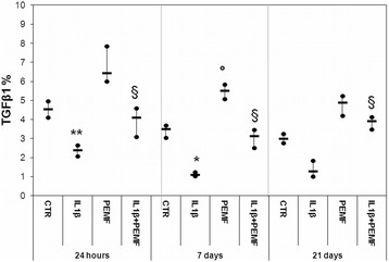
An in vitro OA model was realized, culturing bovine cartilage explants with a high dose of interleukin 1β (IL1β, 50 ng/ml) at different experimental times (24 h, and 7 and 21 days). The effects of PEMFs (75 Hz, 1.5 mT) were evaluated in cartilage explants treated with IL1β or not (control), in terms of cartilage structure, cellularity and proteoglycans, glycosaminoglycans, collagen II and transforming growth factor β1 synthesis by using histology, histomorphometry and immunohistochemistry.
Making a comparison with control cartilage, IL1β-treated explants showed a decrease in cartilage matrix, structure and cellularity parameters. PEMFs were able to counteract the progression of OA acting on both cartilage cellularity and ECM in cartilage previously treated with IL1β. Normal distribution (Kolmogroc-Smirnov test) and homoscedasticity (Levene test) of data were verified, then, the non-parametric Kruskal Wallis test followed by Mann-Whiteny U test for pairwise comparisons were performed. The p-value was adjusted according to the Dunn-Sidak correction.
These results, obtained by culturing and treating cartilage explants from two different joints, confirmed that PEMF stimulation can be used as adjuvant therapy to preserve cartilage from detrimental effects of high inflammatory cytokine levels during OA.
骨关节炎(OA)是关节软骨结构、成分和细胞数量发生渐进性改变的最终结果,随后关节滑液中促炎细胞因子浓度升高。尽管脉冲电磁场(PEMF)刺激因其合成代谢和抗炎特性在对抗OA进展和炎症方面的作用越来越受到关注,但本研究旨在从组织学和组织形态计量学角度,增进对与PEMF暴露相关的软骨细胞外基质(ECM)和软骨细胞变化的认识。
建立体外OA模型,在不同实验时间(24小时、7天和21天)用高剂量白细胞介素1β(IL1β,50 ng/ml)培养牛软骨外植体。通过组织学、组织形态计量学和免疫组织化学,评估PEMF(75 Hz,1.5 mT)对用或未用IL1β处理的软骨外植体(对照)的软骨结构、细胞数量以及蛋白聚糖、糖胺聚糖、胶原蛋白II和转化生长因子β1合成的影响。
与对照软骨相比,用IL1β处理的外植体显示软骨基质、结构和细胞数量参数下降。PEMF能够通过作用于先前用IL1β处理的软骨的细胞数量和ECM来对抗OA的进展。验证了数据的正态分布(Kolmogroc-Smirnov检验)和同方差性(Levene检验),然后进行非参数Kruskal Wallis检验,随后进行Mann-Whiteny U检验进行成对比较。根据Dunn-Sidak校正调整p值。
通过培养和处理来自两个不同关节的软骨外植体获得的这些结果证实,PEMF刺激可作为辅助治疗,保护软骨免受OA期间高炎症细胞因子水平的有害影响。