Ladefoged Claes Nøhr, Hansen Adam Espe, Keller Sune Høgild, Holm Søren, Law Ian, Beyer Thomas, Højgaard Liselotte, Kjær Andreas, Andersen Flemming Littrup
Department of Clinical Physiology, Nuclear Medicine and PET, Rigshospitalet, University of Copenhagen, Blegdamsvej 9, 2100, Copenhagen, Denmark.
Centre for Medical Physics and Biomedical Engineering, Medical University of Vienna, Waehringer Guertel 18-20/4L, Vienna, A-1090, Austria.
EJNMMI Phys. 2014 Dec;1(1):101. doi: 10.1186/s40658-014-0101-0. Epub 2014 Dec 14.
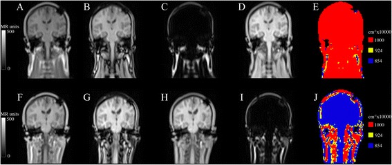
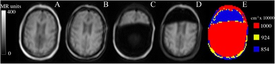
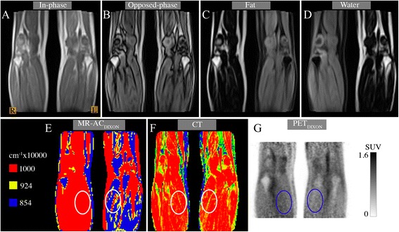
The current MR-based attenuation correction (AC) used in combined PET/MR systems computes a Dixon attenuation map (MR-ACDixon) based on fat and water images derived from in- and opposed-phase MRI. We observed an occasional fat/water inversion in MR-ACDixon. The aim of our study was to estimate the prevalence of this phenomenon in a large patient cohort and assess the possible bias on PET data.
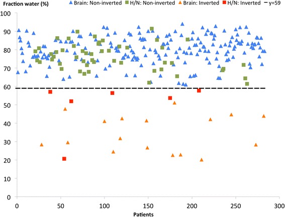
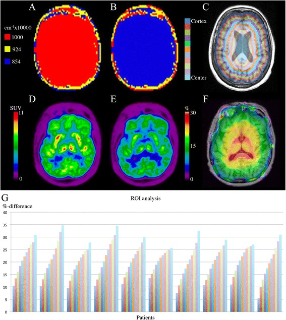
PET/MRI was performed on a Siemens Biograph mMR (Siemens AG, Erlangen, Germany). We visually inspected attenuation maps of 283 brain or head/neck (H/N) patients, classified them as non-inverted or inverted, and calculated the fat/water tissue fraction. We selected ten FDG-PET brain patients with non-inverted attenuation maps for further analysis. Tissue inversion was simulated, and PET images were reconstructed using both original and inverted attenuation maps. The FDG-PET images of the ten brain patients were analyzed using 11 concentric annulus regions of 5 mm width placed over a central transaxial image plane traversing PETDixon.
Out of the 283 patients, a fat/water inversion in 23 patients (8.1%) was observed. The average fraction of fat in the correct MR-ACDixon was 13% for brain and 17% for H/N patients. In the inverted cases, we found an average fat fraction of 56% for the brain patients and 41% for the H/N patients. The effect of the simulated tissue inversion in the brain studies was clearly seen on AC-PET images. The percent-difference image revealed a radial error where the largest difference was at the ventricles (30% ± 3%) and smallest at the cortical region (10% ± 2%).
Tissue inversion in Dixon MRI is well known and can occur when there is an error in the off-resonance correction method. Tissue inversion needs to be considered if, based on Dixon-AC, the construction of normal PET databases is performed or any quantitative physiological parameters are fitted. Visual inspection is needed if Dixon-AC is to be used in clinical routine.
当前在PET/MR联合系统中使用的基于磁共振成像(MR)的衰减校正(AC),根据同相和反相MRI获得的脂肪和水图像计算狄克逊衰减图(MR-ACDixon)。我们观察到MR-ACDixon中偶尔会出现脂肪/水反转现象。本研究的目的是估计在一大群患者中这种现象的发生率,并评估对PET数据可能产生的偏差。
在西门子Biograph mMR(德国埃尔朗根西门子公司)上进行PET/MRI检查。我们对283例脑部或头颈部(H/N)患者的衰减图进行了视觉检查,将其分为未反转或反转,并计算了脂肪/水组织分数。我们选择了10例衰减图未反转的FDG-PET脑部患者进行进一步分析。模拟组织反转,并使用原始和反转的衰减图重建PET图像。使用放置在穿过PETDixon的中央横轴图像平面上的11个宽度为5 mm的同心环形区域,对这10例脑部患者的FDG-PET图像进行分析。
在283例患者中,观察到23例(8.1%)出现脂肪/水反转。在正确的MR-ACDixon中,脑部患者的平均脂肪分数为13%,H/N患者为17%。在反转的病例中,我们发现脑部患者的平均脂肪分数为56%,H/N患者为41%。在脑部研究中,模拟组织反转对AC-PET图像的影响清晰可见。百分比差异图像显示出径向误差,其中最大差异出现在脑室(30%±3%),最小差异出现在皮质区域(10%±2%)。
狄克逊MRI中的组织反转是众所周知的,当失谐校正方法出现错误时可能会发生。如果基于狄克逊AC构建正常PET数据库或拟合任何定量生理参数,则需要考虑组织反转。如果要在临床常规中使用狄克逊AC,则需要进行视觉检查。