Konno Tasuku, Pinho Melo Eduardo, Lopes Carlos, Mehmeti Ilir, Lenzen Sigurd, Ron David, Avezov Edward
University of Cambridge, Cambridge Institute for Medical Research, Wellcome Trust Medical Research Council Institute of Metabolic Science and National Institute for Health Research Cambridge Biomedical Research Centre, Cambridge, CB2 0XY, UK.
Center for Biomedical Research, Universidade do Algarve, Faro, Portugal 8005-139.
J Cell Biol. 2015 Oct 26;211(2):253-9. doi: 10.1083/jcb.201506123.
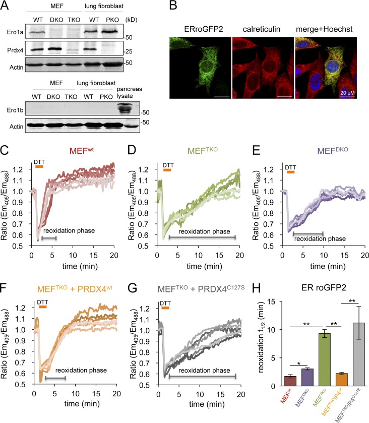
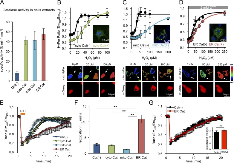
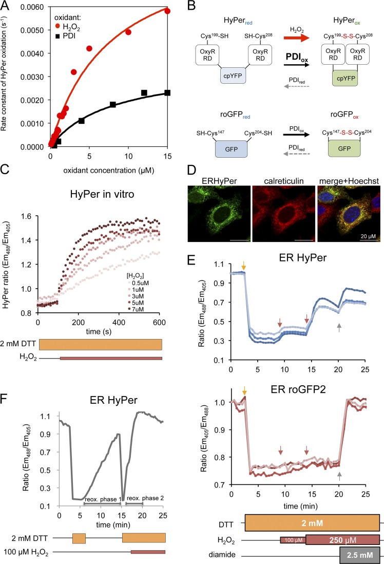
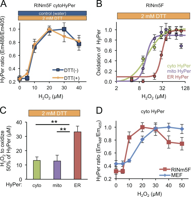
The endoplasmic reticulum (ER)-localized peroxiredoxin 4 (PRDX4) supports disulfide bond formation in eukaryotic cells lacking endoplasmic reticulum oxidase 1 (ERO1). The source of peroxide that fuels PRDX4-mediated disulfide bond formation has remained a mystery, because ERO1 is believed to be a major producer of hydrogen peroxide (H2O2) in the ER lumen. We report on a simple kinetic technique to track H2O2 equilibration between cellular compartments, suggesting that the ER is relatively isolated from cytosolic or mitochondrial H2O2 pools. Furthermore, expression of an ER-adapted catalase to degrade lumenal H2O2 attenuated PRDX4-mediated disulfide bond formation in cells lacking ERO1, whereas depletion of H2O2 in the cytosol or mitochondria had no similar effect. ER catalase did not effect the slow residual disulfide bond formation in cells lacking both ERO1 and PRDX4. These observations point to exploitation of a hitherto unrecognized lumenal source of H2O2 by PRDX4 and a parallel slow H2O2-independent pathway for disulfide formation.
内质网(ER)定位的过氧化物酶4(PRDX4)在缺乏内质网氧化酶1(ERO1)的真核细胞中支持二硫键的形成。为PRDX4介导的二硫键形成提供过氧化氢的来源一直是个谜,因为ERO1被认为是内质网腔中过氧化氢(H2O2)的主要产生者。我们报告了一种简单的动力学技术来追踪细胞区室之间的H2O2平衡,这表明内质网与胞质或线粒体的H2O2池相对隔离。此外,表达一种内质网适应性过氧化氢酶以降解内质网腔中的H2O2,会减弱缺乏ERO1的细胞中PRDX4介导的二硫键形成,而胞质或线粒体中H2O2的消耗则没有类似效果。内质网过氧化氢酶对缺乏ERO1和PRDX4的细胞中缓慢的残余二硫键形成没有影响。这些观察结果表明,PRDX4利用了一种迄今未被认识的内质网腔H2O2来源,以及一条平行的、与H2O2无关的缓慢二硫键形成途径。