Department of Diagnostic and Interventional Imaging, KK Women's and Children's Hospital, Singapore.
Ultrasonography. 2016 Jan;35(1):13-24. doi: 10.14366/usg.15043. Epub 2015 Sep 25.
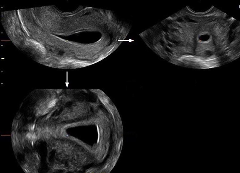
Ultrasonography (US) is the most recent cross-sectional imaging modality to acquire three-dimensional (3D) capabilities. The reconstruction of volumetric US data for multiplanar display took a significantly longer time to develop in comparison with computed tomography and magnetic resonance imaging. The current equipment for 3D-US is capable of producing high-resolution images in three different planes, including real-time surface-rendered images. The use of 3D-US in gynaecology was accelerated through the development of the endovaginal volume transducer, which allows the automated acquisition of volumetric US data. Although initially considered an adjunct to two-dimensional US, 3D-US is now the imaging modality of choice for the assessment of Müllerian duct anomalies and the location of intrauterine devices.
超声检查(US)是最新的三维(3D)成像方式。与计算机断层扫描和磁共振成像相比,三维超声数据的多平面显示重建花费了更长的时间。目前的三维超声设备能够在三个不同的平面上生成高分辨率的图像,包括实时表面渲染图像。经阴道容积探头的发展加速了三维超声在妇科中的应用,该探头可自动采集容积超声数据。虽然三维超声最初被认为是二维超声的辅助手段,但现在它已成为评估苗勒管异常和宫内节育器位置的首选成像方式。