Chen Ya-Wen, Liou Gunn-Guang, Pan Huay-Ben, Tseng Hui-Hwa, Hung Yu-Ting, Chou Chen-Pin
National Institute of Cancer Research, National Health Research Institutes, Miaoli, Taiwan ; Graduate Institute of Basic Medical Science, China Medical University, Taichung, Taiwan.
Institute of Molecular and Genomic Medicine, National Health Research Institutes, Miaoli, Taiwan.
Int J Nanomedicine. 2015 Nov 11;10:6997-7018. doi: 10.2147/IJN.S86592. eCollection 2015.
The use of ultrasmall superparamagnetic iron oxide (USPIO) nanoparticles to visualize cells has been applied clinically, showing the potential for monitoring cells in vivo with magnetic resonance imaging (MRI). USPIO conjugated with anti-CD133 antibodies (USPIO-CD133 Ab) that recognize the CD133 molecule, a cancer stem cell marker in a variety of cancers, was studied as a novel and potent agent for MRI contrast enhancement of tumor cells.
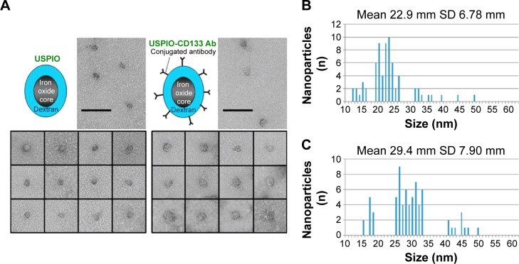
Anti-CD133 antibodies were used to conjugate with USPIO via interaction of streptavidin and biotin for in vivo labeling of CD133-positive cells in xenografted tumors and N-ethyl-N-nitrosourea (ENU)-induced brain tumors. The specific binding of USPIO-CD133 Ab to CD133-positive tumor cells was subsequently detected by Prussian blue staining and MRI with T2-weighted, gradient echo and multiple echo recombined gradient echo images. In addition, the cellular toxicity of USPIO-CD133 Ab was determined by analyzing cell proliferation, apoptosis, and reactive oxygen species production.
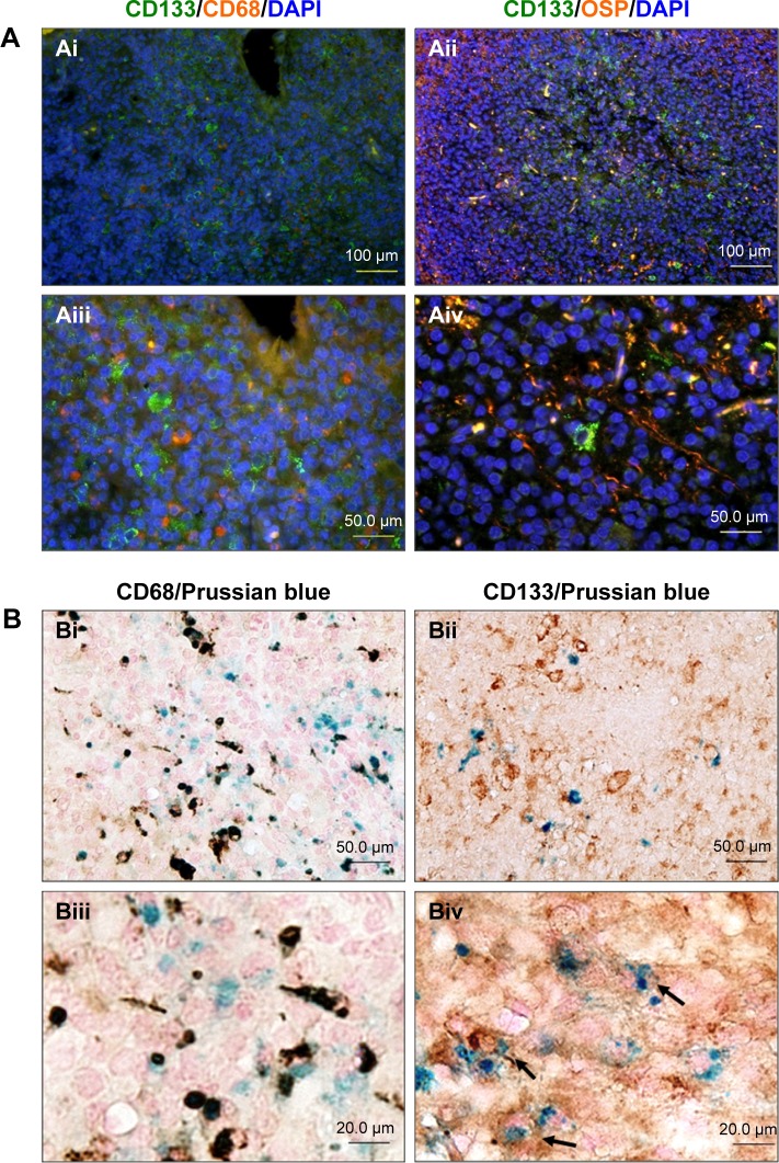
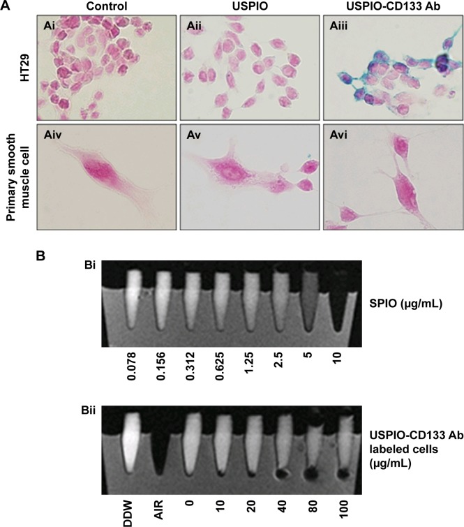
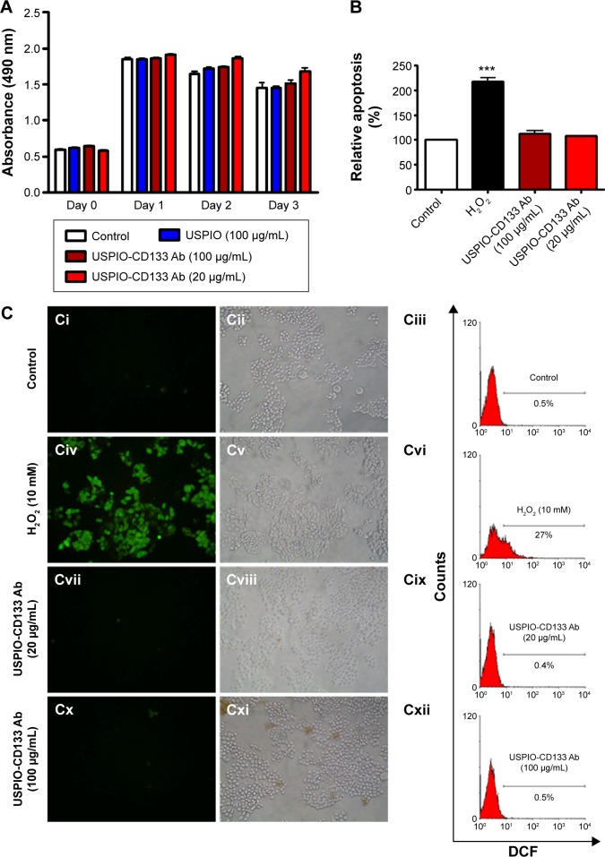
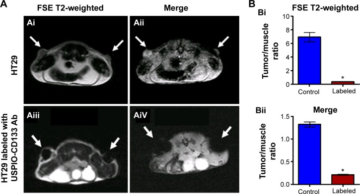
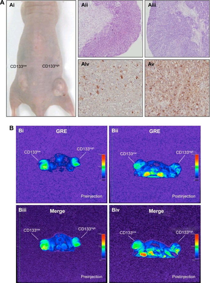
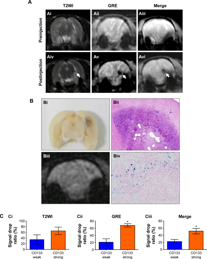
USPIO-CD133 Ab specifically recognizes in vitro and labels CD133-positive cells, as validated using Prussian blue staining and MRI. The assays of cell proliferation, apoptosis, and reactive oxygen species production showed no significant differences in tumor cells with or without labeling of USPIO-CD133 Ab. In vivo imaging of CD133-positive cells was demonstrated by intravenous injection of USPIO-CD133 Ab in mice with HT29 xenografted tumors. The MRI of HT29 xenografts showed several clusters of hypotensive regions that correlated with CD133 expression and Prussian blue staining for iron. In rat, brain tumors induced by transplacental ENU mutagenesis, several clusters of hypointensive zones were observed in CD133-expressing brain tumors by MRI and intravenously administered USPIO-CD133 Ab.
Combination of USPIO-CD133 Ab and MRI is valuable in recognizing CD133-expressing tumor cells in vitro, extracellularly labeling for cell tracking and detecting CD133-expressing tumors in xenografted tumors as well as ENU-induced rat brain tumors.
超小超顺磁性氧化铁(USPIO)纳米颗粒用于细胞可视化已应用于临床,显示出磁共振成像(MRI)在体内监测细胞的潜力。研究了与抗CD133抗体(USPIO-CD133 Ab)偶联的USPIO,该抗体可识别CD133分子,这是多种癌症中的一种癌症干细胞标志物,作为一种新型强效剂用于肿瘤细胞的MRI对比增强。
通过链霉亲和素和生物素的相互作用,使用抗CD133抗体与USPIO偶联,用于体内标记异种移植肿瘤和N-乙基-N-亚硝基脲(ENU)诱导的脑肿瘤中的CD133阳性细胞。随后通过普鲁士蓝染色和T2加权、梯度回波和多回波重组梯度回波图像的MRI检测USPIO-CD133 Ab与CD133阳性肿瘤细胞的特异性结合。此外,通过分析细胞增殖、凋亡和活性氧产生来确定USPIO-CD133 Ab的细胞毒性。
如使用普鲁士蓝染色和MRI所验证的,USPIO-CD133 Ab在体外特异性识别并标记CD133阳性细胞。细胞增殖、凋亡和活性氧产生的测定表明,标记或未标记USPIO-CD133 Ab的肿瘤细胞之间没有显著差异。在携带HT29异种移植肿瘤的小鼠中静脉注射USPIO-CD133 Ab,证明了CD133阳性细胞的体内成像。HT29异种移植瘤的MRI显示出几簇与CD133表达和铁的普鲁士蓝染色相关的低信号区。在大鼠中,经胎盘ENU诱变诱导的脑肿瘤,通过MRI和静脉注射USPIO-CD133 Ab,在表达CD133的脑肿瘤中观察到几簇低信号区。
USPIO-CD133 Ab与MRI的组合在体外识别表达CD133的肿瘤细胞(用于细胞追踪的细胞外标记)以及检测异种移植肿瘤和ENU诱导的大鼠脑肿瘤中表达CD133的肿瘤方面具有重要价值。