Sun Hua Yu, Goodkin Howard P
Department of Neurology, University of Virginia Health System, Charlottesville, VA 22908, USA.
Department of Neurology, University of Virginia Health System, Charlottesville, VA 22908, USA; Department of Pediatrics, University of Virginia Health System, Charlottesville, VA 22908, USA.
Epilepsy Res. 2016 Jan;119:30-3. doi: 10.1016/j.eplepsyres.2015.11.006. Epub 2015 Nov 12.
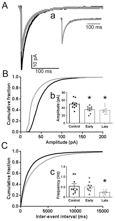
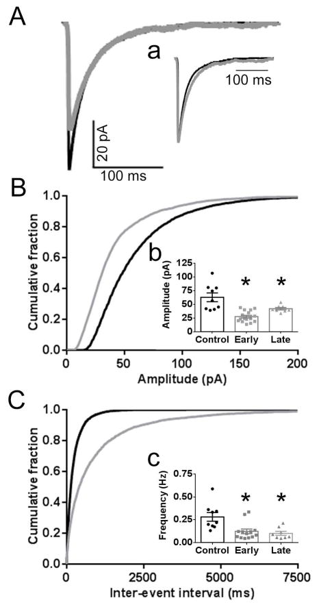
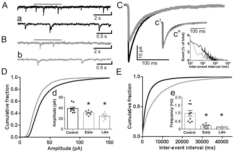
The goal of this study was to determine whether there are region-specific or time-dependent changes in GABA-mediated synaptic inhibition of principal neurons in the hippocampus during in vivo status epilepticus. Standard whole cell patch clamp electrophysiological techniques were used to characterize miniature inhibitory postsynaptic currents (mIPSCs) in recordings from the principal neurons (PNs) of the dentate gyrus, CA1, and CA3 in acutely-obtained hippocampal slices from control and lithium/pilocarpine-induced status epilepticus(SE)-treated animals. The reduction in mIPSC amplitude was pervasive across the 3 regions examined in hippocampal slices obtained after 60 min (late) or just 15 min after the onset of SE. The mIPSC frequency was reduced in all 3 regions after 60 min and 2 regions (dentate, CA1) after 15 min. These findings lend further support to the hypothesis that a rapid modification of the postsynaptic GABAA receptor population leads to a widespread decline in GABA-mediated inhibition that, in part, contributes to both the self-sustaining nature of SE and to the decrease in the efficacy of benzodiazepines.
本研究的目的是确定在体内癫痫持续状态期间,海马体中主要神经元的GABA介导的突触抑制是否存在区域特异性或时间依赖性变化。使用标准的全细胞膜片钳电生理技术,对来自对照动物以及锂/匹鲁卡品诱导的癫痫持续状态(SE)处理动物的急性获取的海马切片中齿状回、CA1和CA3的主要神经元(PNs)记录中的微小抑制性突触后电流(mIPSCs)进行表征。在SE发作60分钟后(晚期)或仅15分钟后获得的海马切片中,所检查的3个区域的mIPSC幅度均普遍降低。60分钟后,所有3个区域的mIPSC频率均降低,15分钟后,2个区域(齿状回、CA1)的mIPSC频率降低。这些发现进一步支持了以下假设:突触后GABAA受体群体的快速改变导致GABA介导的抑制广泛下降,这在一定程度上促成了SE的自我维持特性以及苯二氮䓬类药物疗效的降低。