Greve Gabriele, Schiffmann Insa, Pfeifer Dietmar, Pantic Milena, Schüler Julia, Lübbert Michael
University of Freiburg Medical Center, Freiburg, Germany.
University of Freiburg, Faculty of Biology, Freiburg, Germany.
BMC Cancer. 2015 Dec 16;15:947. doi: 10.1186/s12885-015-1967-5.
The receptor tyrosine kinase (RTK) EGFR is overexpressed and mutated in NSCLC. These mutations can be targeted by RTK inhibitors (TKIs) such as erlotinib. Chromatin-modifying agents may offer a novel therapeutic approach by sensitizing tumor cells to TKIs.
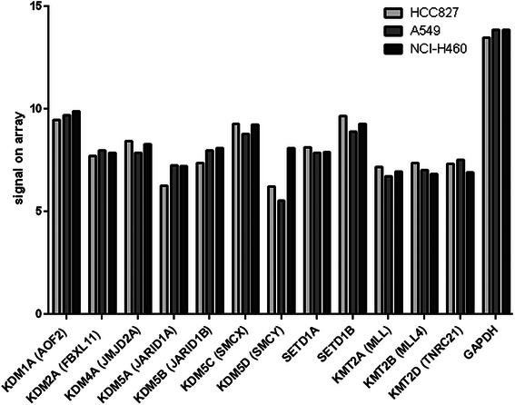
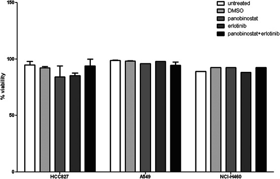
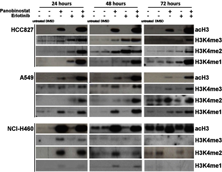
The NSCLC cell lines HCC827 (EGFR mutant, adenocarcinoma), A549 (EGFR wt, adenocarcinoma) and NCI-H460 (EGFR wt, large cell carcinoma) were analyzed by SNP6.0 array. Changes in proliferation after panobinostat (LBH-589, PS) and erlotinib treatment were quantified by WST-1 assay and apoptosis by Annexin V/7-AAD flow cytometry. Abundance of target proteins and histone marks (acH3, H3K4me1/2/3) was determined by immunoblotting.
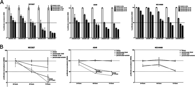
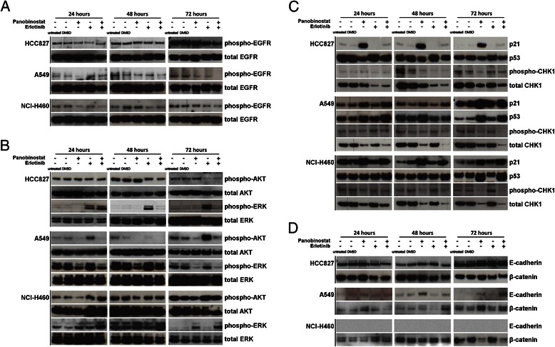
As expected, the EGFR wt cell lines A549 and NCI-H460 were quite insensitive to the growth-inhibitory effect of erlotinib (IC50 70-100 μM), compared to HCC827 (IC50<0.02 μM). All three cell lines were sensitive to PS treatment (IC50: HCC827 10 nM, A549 20 nM and NCI-H460 35 nM). The combination of both drugs further reduced proliferation in HCC827 and in A549, but not in NCI-H460. PS alone induced differentiation and expression of p21WAF1/CIP1 and p53 and decreased CHK1 in all three cell lines, with almost no further effect when combined with erlotinib. In contrast, combination treatment additively decreased pEGFR, pERK and pAKT in A549. Both drugs synergistically induced acH3 in the adenocarcinoma lines. Surprisingly, we also observed induction of H3K4 methylation marks after erlotinib treatment in HCC827 and in A549 that was further enhanced by combination with PS.
PS sensitized lung adenocarcinoma cells to the antiproliferative effects of erlotinib. In these cell lines, the drug combination also had a robust, not previously described effect on histone H3 acetylation and H3K4 methylation.
受体酪氨酸激酶(RTK)表皮生长因子受体(EGFR)在非小细胞肺癌(NSCLC)中过表达且发生突变。这些突变可被如厄洛替尼等RTK抑制剂(TKIs)靶向作用。染色质修饰剂可能通过使肿瘤细胞对TKIs敏感而提供一种新的治疗方法。
采用SNP6.0芯片分析NSCLC细胞系HCC827(EGFR突变型,腺癌)、A549(EGFR野生型,腺癌)和NCI-H460(EGFR野生型,大细胞癌)。通过WST-1法对帕比司他(LBH-589,PS)和厄洛替尼处理后的细胞增殖变化进行定量分析,采用Annexin V/7-AAD流式细胞术检测细胞凋亡情况。通过免疫印迹法测定靶蛋白和组蛋白标记(乙酰化组蛋白H3,H3K4me1/2/3)的丰度。
正如预期,与HCC827(IC50<0.02 μM)相比,EGFR野生型细胞系A549和NCI-H460对厄洛替尼的生长抑制作用相当不敏感(IC50为70 - 100 μM)。所有三种细胞系对PS处理均敏感(IC50:HCC827为10 nM,A549为20 nM,NCI-H460为35 nM)。两种药物联合使用进一步降低了HCC827和A549细胞的增殖,但对NCI-H460细胞无此作用。单独使用PS可诱导所有三种细胞系分化并表达p21WAF1/CIP1和p53,降低CHK1表达,与厄洛替尼联合使用时几乎无进一步影响。相反,联合治疗可使A549细胞中的磷酸化EGFR、磷酸化ERK和磷酸化AKT呈相加性降低。两种药物在腺癌细胞系中协同诱导乙酰化组蛋白H3。令人惊讶的是,我们还观察到厄洛替尼处理后HCC827和A549细胞中H3K4甲基化标记的诱导,与PS联合使用时进一步增强。
PS使肺腺癌细胞对厄洛替尼的抗增殖作用敏感。在这些细胞系中,药物联合使用对组蛋白H3乙酰化和H3K4甲基化也有强大的、此前未描述的作用。