• 文献检索
  • 文档翻译
  • 深度研究
  • 学术资讯
  • Suppr Zotero 插件Zotero 插件
  • 邀请有礼
  • 套餐&价格
  • 历史记录
应用&插件
Suppr Zotero 插件Zotero 插件浏览器插件Mac 客户端Windows 客户端微信小程序
定价
高级版会员购买积分包购买API积分包
服务
文献检索文档翻译深度研究API 文档MCP 服务
关于我们
关于 Suppr公司介绍联系我们用户协议隐私条款
关注我们

Suppr 超能文献

核心技术专利:CN118964589B侵权必究
粤ICP备2023148730 号-1Suppr @ 2026

文献检索

告别复杂PubMed语法,用中文像聊天一样搜索,搜遍4000万医学文献。AI智能推荐,让科研检索更轻松。

立即免费搜索

文件翻译

保留排版,准确专业,支持PDF/Word/PPT等文件格式,支持 12+语言互译。

免费翻译文档

深度研究

AI帮你快速写综述,25分钟生成高质量综述,智能提取关键信息,辅助科研写作。

立即免费体验

用于确定干细胞分化的髓核表型标志物:事实还是虚构?

Nucleus pulposus phenotypic markers to determine stem cell differentiation: fact or fiction?

作者信息

Thorpe Abbey A, Binch Abbie L A, Creemers Laura B, Sammon Christopher, Le Maitre Christine L

机构信息

Biomolecular Sciences Research Centre, Sheffield Hallam University, Sheffield, UK.

UMC Utrecht, Orthopaedics Department, Utrecht, Netherlands.

出版信息

Oncotarget. 2016 Jan 19;7(3):2189-200. doi: 10.18632/oncotarget.6782.

DOI:10.18632/oncotarget.6782
PMID:26735178
原文链接:https://pmc.ncbi.nlm.nih.gov/articles/PMC4823028/
Abstract

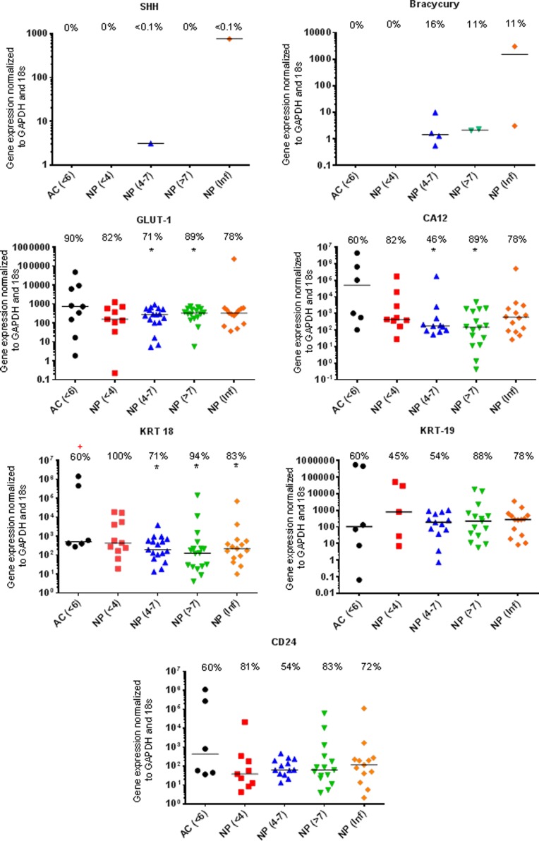
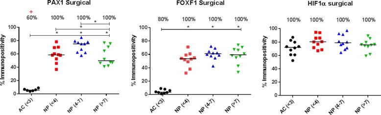
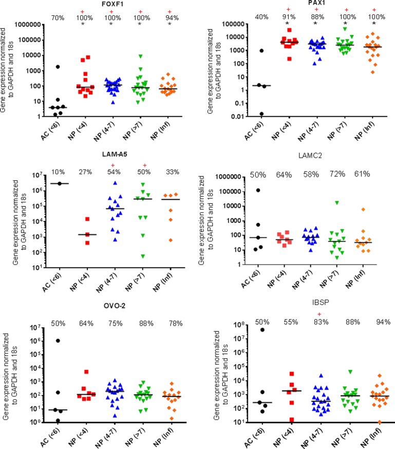
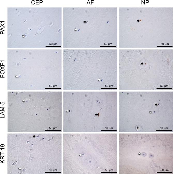
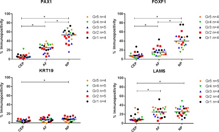
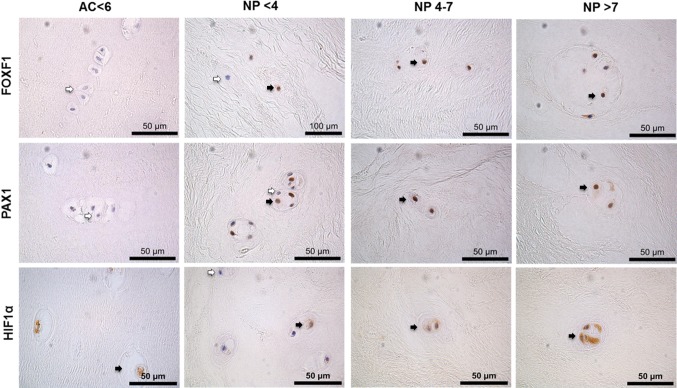
Progress in mesenchymal stem cell (MSC) based therapies for nucleus pulposus (NP) regeneration are hampered by a lack of understanding and consensus of the normal NP cell phenotype. Despite the recent consensus paper on NP markers, there is still a need to further validate proposed markers. This study aimed to determine whether an NP phenotypic profile could be identified within a large population of mature NP samples.qRT-PCR was conducted to assess mRNA expression of 13 genes within human non-degenerate articular chondrocytes (AC) (n=10) and NP cells extracted from patients across a spectrum of histological degeneration grades (n=71). qRT-PCR results were used to select NP marker candidates for protein expression analysis.Differential expression at mRNA between AC and non-degenerate NP cells was only observed for Paired Box Protein 1 (PAX1) and Forkhead box F1 (FOXF1). In contrast no other previously suggested markers displayed differential expression between non-degenerate NP and AC at mRNA level. PAX1 and FOXF1 protein expression was significantly higher in the NP compared to annulus fibrosus (AF), cartilaginous endplate (CEP) and AC. In contrast Laminin-5 (LAM-332), Keratin-19 (KRT-19) and Hypoxia Inducible Factor 1 alpha (HIF1α) showed no differential expression in NP cells compared with AC cells.A marker which exclusively differentiates NP cells from AF and AC cells remains to be identified, raising the question: is the NP a heterogeneous population of cells? Or does the natural biological variation during IVD development, degeneration state and even the life cycle of cells make finding one definitive marker impossible?

摘要

基于间充质干细胞(MSC)的椎间盘髓核(NP)再生疗法的进展因对正常NP细胞表型缺乏理解和共识而受阻。尽管最近有关于NP标志物的共识文件,但仍需要进一步验证所提出的标志物。本研究旨在确定是否能在大量成熟NP样本中识别出NP表型特征。进行qRT-PCR以评估10例人非退变关节软骨细胞(AC)和71例从不同组织学退变等级患者中提取的NP细胞中13个基因的mRNA表达。qRT-PCR结果用于选择NP标志物候选物进行蛋白质表达分析。仅在配对盒蛋白1(PAX1)和叉头框F1(FOXF1)方面观察到AC和非退变NP细胞之间mRNA的差异表达。相比之下,之前提出的其他标志物在非退变NP和AC之间的mRNA水平上未显示差异表达。与纤维环(AF)、软骨终板(CEP)和AC相比,NP中PAX1和FOXF1蛋白表达显著更高。相比之下,层粘连蛋白-5(LAM-332)、角蛋白-19(KRT-19)和缺氧诱导因子1α(HIF1α)在NP细胞与AC细胞中未显示差异表达。一种能将NP细胞与AF和AC细胞完全区分开的标志物仍有待确定,这就引发了一个问题:NP是细胞的异质群体吗?还是椎间盘退变发展过程中、退变状态甚至细胞生命周期中的自然生物学变异使得找到一个确定的标志物成为不可能?

https://cdn.ncbi.nlm.nih.gov/pmc/blobs/32a4/4823028/d79a9b881514/oncotarget-07-2189-g006.jpg
https://cdn.ncbi.nlm.nih.gov/pmc/blobs/32a4/4823028/1b7ff7031e0c/oncotarget-07-2189-g001.jpg
https://cdn.ncbi.nlm.nih.gov/pmc/blobs/32a4/4823028/0d49cd0396fa/oncotarget-07-2189-g002.jpg
https://cdn.ncbi.nlm.nih.gov/pmc/blobs/32a4/4823028/6d84e0a1d7a2/oncotarget-07-2189-g003.jpg
https://cdn.ncbi.nlm.nih.gov/pmc/blobs/32a4/4823028/90a0fa52510c/oncotarget-07-2189-g004.jpg
https://cdn.ncbi.nlm.nih.gov/pmc/blobs/32a4/4823028/30d4b0415ed1/oncotarget-07-2189-g005.jpg
https://cdn.ncbi.nlm.nih.gov/pmc/blobs/32a4/4823028/d79a9b881514/oncotarget-07-2189-g006.jpg

相似文献

1
Nucleus pulposus phenotypic markers to determine stem cell differentiation: fact or fiction?用于确定干细胞分化的髓核表型标志物:事实还是虚构?
Oncotarget. 2016 Jan 19;7(3):2189-200. doi: 10.18632/oncotarget.6782.
2
Characterization of the human nucleus pulposus cell phenotype and evaluation of novel marker gene expression to define adult stem cell differentiation.人髓核细胞表型的特征分析及新型标记基因表达的评估以界定成体干细胞分化
Arthritis Rheum. 2010 Dec;62(12):3695-705. doi: 10.1002/art.27710.
3
Transcriptional profiling of bovine intervertebral disc cells: implications for identification of normal and degenerate human intervertebral disc cell phenotypes.牛椎间盘细胞的转录组分析:对鉴定正常和退变人类椎间盘细胞表型的启示。
Arthritis Res Ther. 2010;12(1):R22. doi: 10.1186/ar2929. Epub 2010 Feb 11.
4
In search of nucleus pulposus-specific molecular markers.寻找椎间盘特异性分子标志物。
Rheumatology (Oxford). 2014 Apr;53(4):600-10. doi: 10.1093/rheumatology/ket303. Epub 2013 Sep 18.
5
A phenotypic comparison of intervertebral disc and articular cartilage cells in the rat.大鼠椎间盘与关节软骨细胞的表型比较
Eur Spine J. 2007 Dec;16(12):2174-85. doi: 10.1007/s00586-007-0475-y. Epub 2007 Sep 5.
6
Differentiation of menstrual blood-derived stem cells toward nucleus pulposus-like cells in a coculture system with nucleus pulposus cells.在与髓核细胞的共培养系统中,月经血源性干细胞向髓核样细胞的分化。
Spine (Phila Pa 1976). 2014 Apr 20;39(9):754-60. doi: 10.1097/BRS.0000000000000261.
7
Variations in gene and protein expression in human nucleus pulposus in comparison with annulus fibrosus and cartilage cells: potential associations with aging and degeneration.人髓核组织中基因和蛋白表达的变化与纤维环和软骨细胞的比较:与衰老和退变的潜在关联。
Osteoarthritis Cartilage. 2010 Mar;18(3):416-23. doi: 10.1016/j.joca.2009.09.009. Epub 2009 Oct 1.
8
Isolation and characterization of mesenchymal stromal cells from human degenerated nucleus pulposus: comparison with bone marrow mesenchymal stromal cells from the same subjects.人退变髓核组织中间充质基质细胞的分离与鉴定:与同一供体来源骨髓间充质基质细胞的比较。
Spine (Phila Pa 1976). 2010 Dec 15;35(26):2259-65. doi: 10.1097/BRS.0b013e3181cb8828.
9
Thermally triggered injectable hydrogel, which induces mesenchymal stem cell differentiation to nucleus pulposus cells: Potential for regeneration of the intervertebral disc.热触发可注射水凝胶,可诱导间充质干细胞分化为髓核细胞:椎间盘再生的潜力。
Acta Biomater. 2016 May;36:99-111. doi: 10.1016/j.actbio.2016.03.029. Epub 2016 Mar 17.
10
Novel immortal human cell lines reveal subpopulations in the nucleus pulposus.新型永生人类细胞系揭示了髓核中的亚群。
Arthritis Res Ther. 2014 Jun 27;16(3):R135. doi: 10.1186/ar4597.

引用本文的文献

1
Engineering Intervertebral Disc Regeneration: Biomaterials, Cell Sources and Animal Models.工程化椎间盘再生:生物材料、细胞来源与动物模型
Cell Prolif. 2025 Sep;58(9):e70046. doi: 10.1111/cpr.70046. Epub 2025 May 19.
2
Current Therapeutic Strategies of Intervertebral Disc Regenerative Medicine.当前椎间盘再生医学的治疗策略。
Mol Diagn Ther. 2024 Nov;28(6):745-775. doi: 10.1007/s40291-024-00729-7. Epub 2024 Aug 19.
3
Assessment of Tie2-Rejuvenated Nucleus Pulposus Cell Transplants from Young and Old Patient Sources Demonstrates That Age Still Matters.

本文引用的文献

1
Degeneration of the intervertebral disc with new approaches for treating low back pain.椎间盘退变与治疗腰痛的新方法
J Neurosurg Sci. 2015 Mar;59(1):47-61. Epub 2014 Nov 25.
2
Defining the phenotype of young healthy nucleus pulposus cells: recommendations of the Spine Research Interest Group at the 2014 annual ORS meeting.定义年轻健康髓核细胞的表型:脊柱研究兴趣小组在2014年ORS年会的建议
J Orthop Res. 2015 Mar;33(3):283-93. doi: 10.1002/jor.22789. Epub 2015 Jan 21.
3
Loss of HIF-1α in the notochord results in cell death and complete disappearance of the nucleus pulposus.
评估来自年轻和老年患者来源的 Tie2 再生活体细胞移植,结果表明年龄仍然很重要。
Int J Mol Sci. 2024 Jul 30;25(15):8335. doi: 10.3390/ijms25158335.
4
Getting to the Core: Exploring the Embryonic Development from Notochord to Nucleus Pulposus.深入核心:探索从脊索到髓核的胚胎发育过程。
J Dev Biol. 2024 Jul 3;12(3):18. doi: 10.3390/jdb12030018.
5
Future of low back pain: unravelling IVD components and MSCs' potential.腰痛的未来:解析椎间盘组成部分及间充质干细胞的潜力
Cell Regen. 2024 Jan 16;13(1):1. doi: 10.1186/s13619-023-00184-5.
6
Recommendations for intervertebral disc notochordal cell investigation: From isolation to characterization.椎间盘脊索细胞研究建议:从分离到表征
JOR Spine. 2023 Jul 9;6(3):e1272. doi: 10.1002/jsp2.1272. eCollection 2023 Sep.
7
An Automatized Deep Segmentation and Classification Model for Lumbar Disk Degeneration and Clarification of Its Impact on Clinical Decisions.一种用于腰椎间盘退变的自动化深度分割与分类模型及其对临床决策影响的阐释
Global Spine J. 2025 Mar;15(2):554-563. doi: 10.1177/21925682231200783. Epub 2023 Sep 12.
8
Alterations of bovine nucleus pulposus cells with aging.牛髓核细胞的老化改变。
Aging Cell. 2023 Aug;22(8):e13873. doi: 10.1111/acel.13873. Epub 2023 May 30.
9
and oxaloacetate in nucleus pulposus stromal cell somatic reprogramming and survival.以及草酰乙酸在髓核基质细胞体细胞重编程和存活中的作用。
Front Mol Biosci. 2022 Nov 3;9:1009402. doi: 10.3389/fmolb.2022.1009402. eCollection 2022.
10
Notochordal Cell-Based Treatment Strategies and Their Potential in Intervertebral Disc Regeneration.基于脊索细胞的治疗策略及其在椎间盘再生中的潜力。
Front Cell Dev Biol. 2022 Mar 14;9:780749. doi: 10.3389/fcell.2021.780749. eCollection 2021.
脊索中缺氧诱导因子-1α的缺失导致细胞死亡和髓核完全消失。
PLoS One. 2014 Oct 22;9(10):e110768. doi: 10.1371/journal.pone.0110768. eCollection 2014.
4
Novel immortal human cell lines reveal subpopulations in the nucleus pulposus.新型永生人类细胞系揭示了髓核中的亚群。
Arthritis Res Ther. 2014 Jun 27;16(3):R135. doi: 10.1186/ar4597.
5
Reflecting on the global burden of musculoskeletal conditions: lessons learnt from the global burden of disease 2010 study and the next steps forward.反思肌肉骨骼疾病的全球负担:从 2010 年全球疾病负担研究中吸取的教训及未来的发展方向。
Ann Rheum Dis. 2015 Jan;74(1):4-7. doi: 10.1136/annrheumdis-2014-205393. Epub 2014 Jun 9.
6
Wnt signaling activates Shh signaling in early postnatal intervertebral discs, and re-activates Shh signaling in old discs in the mouse.Wnt 信号在新生后椎间盘早期激活 Shh 信号,在小鼠老年椎间盘中重新激活 Shh 信号。
PLoS One. 2014 Jun 3;9(6):e98444. doi: 10.1371/journal.pone.0098444. eCollection 2014.
7
Cannabinoid WIN-55,212-2 mesylate inhibits interleukin-1β induced matrix metalloproteinase and tissue inhibitor of matrix metalloproteinase expression in human chondrocytes.甲磺酸大麻隆 WIN-55,212-2 抑制白细胞介素-1β诱导的人软骨细胞基质金属蛋白酶和基质金属蛋白酶组织抑制剂的表达。
Osteoarthritis Cartilage. 2014 Jan;22(1):133-44. doi: 10.1016/j.joca.2013.10.016. Epub 2013 Nov 6.
8
Identification of novel nucleus pulposus markers: Interspecies variations and implications for cell-based therapiesfor intervertebral disc degeneration.鉴定新的髓核标志物:种间差异及其对椎间盘退变的基于细胞治疗的意义。
Bone Joint Res. 2013 Aug 19;2(8):169-78. doi: 10.1302/2046-3758.28.2000184. Print 2013.
9
Integrin-mediated interactions with extracellular matrix proteins for nucleus pulposus cells of the human intervertebral disc.整合素介导的细胞外基质蛋白与人类椎间盘髓核细胞的相互作用。
J Orthop Res. 2013 Oct;31(10):1661-7. doi: 10.1002/jor.22395. Epub 2013 Jun 4.
10
Inflammatory cytokines induce NOTCH signaling in nucleus pulposus cells: implications in intervertebral disc degeneration.炎性细胞因子诱导髓核细胞中的 NOTCH 信号通路:在椎间盘退变中的意义。
J Biol Chem. 2013 Jun 7;288(23):16761-16774. doi: 10.1074/jbc.M112.446633. Epub 2013 Apr 15.