Handel Adam E, Chintawar Satyan, Lalic Tatjana, Whiteley Emma, Vowles Jane, Giustacchini Alice, Argoud Karene, Sopp Paul, Nakanishi Mahito, Bowden Rory, Cowley Sally, Newey Sarah, Akerman Colin, Ponting Chris P, Cader M Zameel
Department of Physiology, Anatomy and Genetics, University of Oxford, Oxford, Oxfordshire OX1 3QX, UK, Weatherall Institute of Molecular Medicine, University of Oxford, Oxford, Oxfordshire OX3 9DS, UK.
Weatherall Institute of Molecular Medicine, University of Oxford, Oxford, Oxfordshire OX3 9DS, UK.
Hum Mol Genet. 2016 Mar 1;25(5):989-1000. doi: 10.1093/hmg/ddv637. Epub 2016 Jan 5.
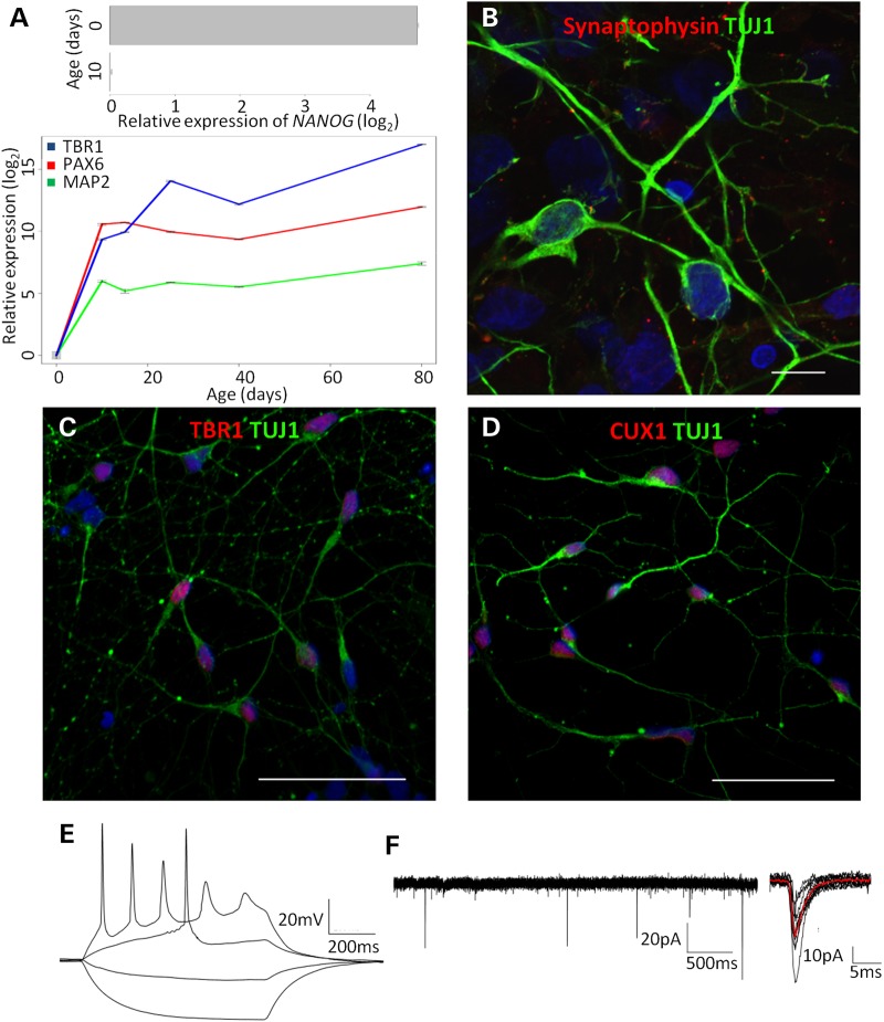
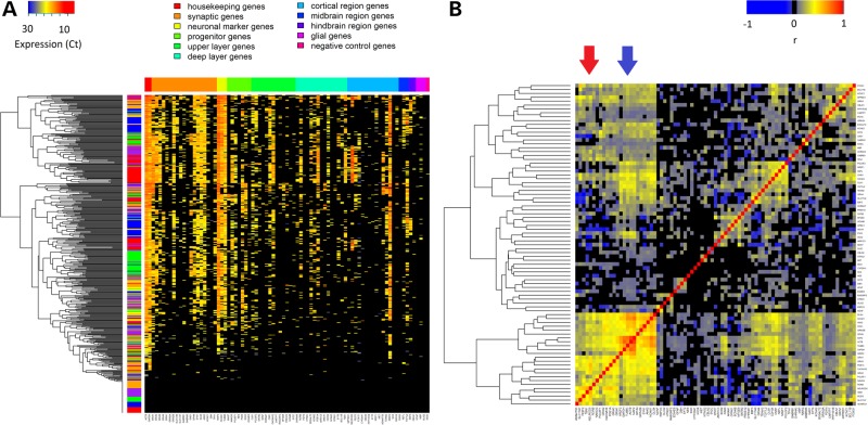
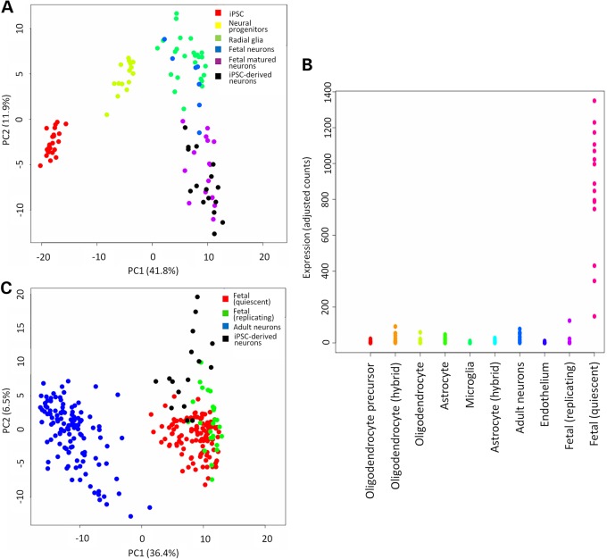
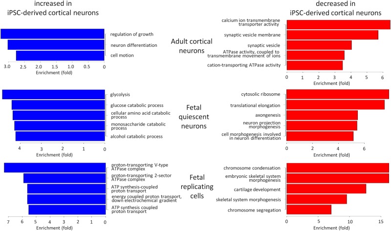
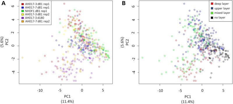
Induced pluripotent stem cell (iPSC)-derived cortical neurons potentially present a powerful new model to understand corticogenesis and neurological disease. Previous work has established that differentiation protocols can produce cortical neurons, but little has been done to characterize these at cellular resolution. In particular, it is unclear to what extent in vitro two-dimensional, relatively disordered culture conditions recapitulate the development of in vivo cortical layer identity. Single-cell multiplex reverse transcriptase-quantitative polymerase chain reaction (RT-qPCR) was used to interrogate the expression of genes previously implicated in cortical layer or phenotypic identity in individual cells. Totally, 93.6% of single cells derived from iPSCs expressed genes indicative of neuronal identity. High proportions of single neurons derived from iPSCs expressed glutamatergic receptors and synaptic genes. And, 68.4% of iPSC-derived neurons expressing at least one layer marker could be assigned to a laminar identity using canonical cortical layer marker genes. We compared single-cell RNA-seq of our iPSC-derived neurons to available single-cell RNA-seq data from human fetal and adult brain and found that iPSC-derived cortical neurons closely resembled primary fetal brain cells. Unexpectedly, a subpopulation of iPSC-derived neurons co-expressed canonical fetal deep and upper cortical layer markers. However, this appeared to be concordant with data from primary cells. Our results therefore provide reassurance that iPSC-derived cortical neurons are highly similar to primary cortical neurons at the level of single cells but suggest that current layer markers, although effective, may not be able to disambiguate cortical layer identity in all cells.
诱导多能干细胞(iPSC)来源的皮质神经元可能是一种强大的新模型,用于理解皮质发生和神经疾病。先前的研究已经证实,分化方案可以产生皮质神经元,但在细胞分辨率水平上对这些神经元进行表征的工作做得很少。特别是,目前尚不清楚体外二维、相对无序的培养条件在多大程度上能够重现体内皮质层身份的发育过程。单细胞多重逆转录酶定量聚合酶链反应(RT-qPCR)被用于检测先前与单个细胞的皮质层或表型身份相关的基因的表达。总体而言,93.6%的iPSC来源的单细胞表达了指示神经元身份的基因。高比例的iPSC来源的单个神经元表达了谷氨酸能受体和突触相关基因。此外,使用经典的皮质层标记基因,68.4%表达至少一种层标记的iPSC来源的神经元可以被确定其层身份。我们将iPSC来源的神经元的单细胞RNA测序结果与来自人类胎儿和成人脑的现有单细胞RNA测序数据进行了比较,发现iPSC来源的皮质神经元与原代胎儿脑细胞非常相似。出乎意料的是,iPSC来源的神经元的一个亚群共同表达了经典的胎儿深层和上层皮质层标记。然而,这似乎与原代细胞的数据一致。因此,我们的结果表明,iPSC来源的皮质神经元在单细胞水平上与原代皮质神经元高度相似,但也表明目前的层标记尽管有效,但可能无法在所有细胞中明确区分皮质层身份。