• 文献检索
  • 文档翻译
  • 深度研究
  • 学术资讯
  • Suppr Zotero 插件Zotero 插件
  • 邀请有礼
  • 套餐&价格
  • 历史记录
应用&插件
Suppr Zotero 插件Zotero 插件浏览器插件Mac 客户端Windows 客户端微信小程序
定价
高级版会员购买积分包购买API积分包
服务
文献检索文档翻译深度研究API 文档MCP 服务
关于我们
关于 Suppr公司介绍联系我们用户协议隐私条款
关注我们

Suppr 超能文献

核心技术专利:CN118964589B侵权必究
粤ICP备2023148730 号-1Suppr @ 2026

文献检索

告别复杂PubMed语法,用中文像聊天一样搜索,搜遍4000万医学文献。AI智能推荐,让科研检索更轻松。

立即免费搜索

文件翻译

保留排版,准确专业,支持PDF/Word/PPT等文件格式,支持 12+语言互译。

免费翻译文档

深度研究

AI帮你快速写综述,25分钟生成高质量综述,智能提取关键信息,辅助科研写作。

立即免费体验

在Descemet膜内皮角膜移植术中两种植片制备方法后内皮细胞活力的逐细胞整体评估。

Global cell-by-cell evaluation of endothelial viability after two methods of graft preparation in Descemet membrane endothelial keratoplasty.

作者信息

Bhogal Maninder, Balda Maria S, Matter Karl, Allan Bruce D

机构信息

Department of Corneal and External Disease, Moorfields Eye Hospital, London, UK Department of Cell Biology, University College London, Institute of Ophthalmology, London, UK.

Department of Cell Biology, University College London, Institute of Ophthalmology, London, UK.

出版信息

Br J Ophthalmol. 2016 Apr;100(4):572-8. doi: 10.1136/bjophthalmol-2015-307534. Epub 2016 Jan 6.

DOI:10.1136/bjophthalmol-2015-307534
PMID:26740609
原文链接:https://pmc.ncbi.nlm.nih.gov/articles/PMC4819631/
Abstract

PURPOSE

To describe a novel method of global cell viability assessment for Descemet membrane endothelial keratoplasty (DMEK) and the comparison of two contemporary methods of donor tissue preparation.

METHODS

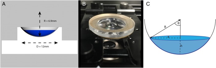
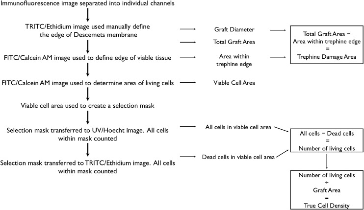
DMEK transplants were prepared using two different methods: liquid bubble separation and manual peeling (n=8 each group). Samples were incubated with Hoechst, calcein-AM and ethidium homodimer prior to mounting on a curved imaging chamber. Z-stacked fluorescence microscopy images were combined to produce an in-focus global image capable of resolving all cell nuclei. Image processing software was used to define a calcein-positive live cell area, count all cell nuclei within this area and subtract ethidium-positive dead cells to derive the total viable endothelial cell count. Corrected global cell density was calculated by dividing the number of viable cells by the graft area, which had been corrected for imaging a curved surface.

RESULTS

Corrected global cell density was lower than the central endothelial cell density in both groups: 85.5% of the pre-preparation central endothelial cell density in the peel group and 75.8% in the bubble group. Corrected global cell density was significantly lower in the liquid bubble separation group than in the peel group (p=0.04).

CONCLUSIONS

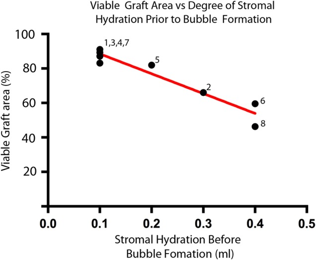
Eye bank estimations of central endothelial cell density overestimate true cell density after graft preparation in DMEK. A peel method is less damaging and more consistent than a liquid bubble method. Cell loss correlated strongly with the degree of stromal hydration prior to bubble separation in the liquid bubble group.

摘要

目的

描述一种用于Descemet膜内皮角膜移植术(DMEK)的新型整体细胞活力评估方法,并比较两种当代供体组织制备方法。

方法

使用两种不同方法制备DMEK移植物:液泡分离法和手工剥离法(每组n = 8)。在将样本安装到弯曲成像室之前,用Hoechst、钙黄绿素-AM和碘化丙啶同型二聚体孵育。将Z轴堆叠的荧光显微镜图像合并,以生成能够分辨所有细胞核的聚焦整体图像。使用图像处理软件定义钙黄绿素阳性活细胞区域,计算该区域内的所有细胞核数量,并减去碘化丙啶阳性死细胞,以得出存活内皮细胞总数。通过将活细胞数量除以已针对弯曲表面成像进行校正的移植物面积,计算校正后的整体细胞密度。

结果

两组校正后的整体细胞密度均低于中央内皮细胞密度:剥离组为制备前中央内皮细胞密度的85.5%,液泡组为75.8%。液泡分离组校正后的整体细胞密度显著低于剥离组(p = 0.04)。

结论

眼库对DMEK移植物制备后中央内皮细胞密度的估计高估了真实细胞密度。与液泡法相比,剥离法的损伤更小且更稳定。在液泡组中,细胞损失与液泡分离前基质水化程度密切相关。

https://cdn.ncbi.nlm.nih.gov/pmc/blobs/afb0/4819631/a361c90eb597/bjophthalmol-2015-307534f05.jpg
https://cdn.ncbi.nlm.nih.gov/pmc/blobs/afb0/4819631/e77de8d30281/bjophthalmol-2015-307534f01.jpg
https://cdn.ncbi.nlm.nih.gov/pmc/blobs/afb0/4819631/6a1a8e021ed1/bjophthalmol-2015-307534f02.jpg
https://cdn.ncbi.nlm.nih.gov/pmc/blobs/afb0/4819631/719186ad6cc8/bjophthalmol-2015-307534f03.jpg
https://cdn.ncbi.nlm.nih.gov/pmc/blobs/afb0/4819631/0156a90701b5/bjophthalmol-2015-307534f04.jpg
https://cdn.ncbi.nlm.nih.gov/pmc/blobs/afb0/4819631/a361c90eb597/bjophthalmol-2015-307534f05.jpg

相似文献

1
Global cell-by-cell evaluation of endothelial viability after two methods of graft preparation in Descemet membrane endothelial keratoplasty.在Descemet膜内皮角膜移植术中两种植片制备方法后内皮细胞活力的逐细胞整体评估。
Br J Ophthalmol. 2016 Apr;100(4):572-8. doi: 10.1136/bjophthalmol-2015-307534. Epub 2016 Jan 6.
2
Endothelial Cell Viability after DMEK Graft Preparation.DMEK移植物制备后的内皮细胞活力
Curr Eye Res. 2021 Nov;46(11):1621-1630. doi: 10.1080/02713683.2021.1927111. Epub 2021 May 24.
3
Ink Retention and Endothelial Cell Viability After the Application of an Orientation Stamp Over an Air Bubble During Descemet Membrane Endothelial Keratoplasty Graft Preparation.在准备Descemet 膜内皮角膜移植术中应用气泡上方的定向印模时的墨水保留和内皮细胞活力。
Cornea. 2021 Jul 1;40(7):894-898. doi: 10.1097/ICO.0000000000002590.
4
Organ culture storage of pre-prepared corneal donor material for Descemet's membrane endothelial keratoplasty.用于Descemet膜内皮角膜移植术的预制备角膜供体材料的器官培养保存
Br J Ophthalmol. 2016 Nov;100(11):1576-1583. doi: 10.1136/bjophthalmol-2016-308855. Epub 2016 Aug 19.
5
Endothelial cell loss following tissue harvesting by pneumodissection for endothelial keratoplasty: an ex vivo study.用于内皮角膜移植术的气动剥离组织采集后内皮细胞损失:一项体外研究。
Br J Ophthalmol. 2015 May;99(5):710-3. doi: 10.1136/bjophthalmol-2014-306560. Epub 2015 Mar 4.
6
Graft preparation for hemi-Descemet membrane endothelial keratoplasty (hemi-DMEK).半厚角膜后弹力层内皮角膜移植术(hemi-DMEK)的植片制备
Br J Ophthalmol. 2016 Mar;100(3):420-4. doi: 10.1136/bjophthalmol-2015-307335. Epub 2015 Oct 27.
7
A comparative study on different Descemet membrane endothelial keratoplasty graft preparation techniques.不同的 Descemet 膜内皮角膜移植术移植物制备技术的比较研究。
Acta Ophthalmol. 2018 Sep;96(6):e718-e726. doi: 10.1111/aos.13746. Epub 2018 Mar 9.
8
Bubble technique for Descemet membrane endothelial keratoplasty tissue preparation in an eye bank: air or liquid?眼库中用于Descemet膜内皮角膜移植术组织制备的气泡技术:空气还是液体?
Acta Ophthalmol. 2015 Mar;93(2):e129-34. doi: 10.1111/aos.12520. Epub 2014 Aug 27.
9
Imaging and quantification of endothelial cell loss in eye bank prepared DMEK grafts using trainable segmentation software.使用可训练分割软件对眼库制备的DMEK移植物中的内皮细胞损失进行成像和定量分析。
Curr Eye Res. 2014 Sep;39(9):894-901. doi: 10.3109/02713683.2014.887120. Epub 2014 Mar 3.
10
24 Effect of graft preparation techniques on clinical outcomes after Descemet membrane endothelial keratoplasty (DMEK).24. 移植制备技术对 Descemet 膜内皮角膜移植术(DMEK)后临床结果的影响。
BMJ Open Ophthalmol. 2022 Nov;7(Suppl 2):A10-A11. doi: 10.1136/bmjophth-2022-EEBA.24.

引用本文的文献

1
Optimized laboratory techniques for assessing the quality of pre-stripped DMEK grafts.评估预剥除DMEK移植物质量的优化实验室技术。
Sci Rep. 2025 Mar 4;15(1):7527. doi: 10.1038/s41598-025-91512-z.
2
Establishment of an Ex Vivo Human Corneal Endothelium Wound Model.体外人角膜内皮细胞损伤模型的建立。
Transl Vis Sci Technol. 2025 Jan 2;14(1):24. doi: 10.1167/tvst.14.1.24.
3
Review of current methods of preparation and distribution of DMEK graft at the eye banks.眼库中角膜后弹力层内皮移植术(DMEK)移植物当前制备与分发方法的综述。

本文引用的文献

1
Evaluation of internationally shipped prestripped donor tissue for descemet membrane endothelial keratoplasty by vital dye staining.通过活体染料染色评估国际运输的预剥离供体组织用于Descemet膜内皮角膜移植术的情况。
Cornea. 2015 Feb;34(2):225-7. doi: 10.1097/ICO.0000000000000330.
2
Air reinjection and endothelial cell density in Descemet membrane endothelial keratoplasty: five-year follow-up.Descemet膜内皮角膜移植术中的空气再注入与内皮细胞密度:五年随访
J Cataract Refract Surg. 2014 Jul;40(7):1116-21. doi: 10.1016/j.jcrs.2014.04.023.
3
Descemet membrane endothelial keratoplasty tissue preparation from donor corneas using a standardized submerged hydro-separation method.
Indian J Ophthalmol. 2025 Jan 1;73(1):19-27. doi: 10.4103/IJO.IJO_243_24. Epub 2024 Dec 23.
4
Descemet Membrane Endothelial Keratoplasty Graft Preparation Using the Liquid Bubble Technique with Subtrabecular Hydrodissection: A Retrospective Real-Life Study.使用带小梁下水分剥离的液泡技术进行Descemet膜内皮角膜移植术的植片制备:一项回顾性真实病例研究。
J Clin Med. 2024 Oct 10;13(20):6048. doi: 10.3390/jcm13206048.
5
Clinical outcome analysis of two approaches to trypan blue dyeing for DMEK.两种 DMEK 中应用台盼蓝染色方法的临床效果分析。
Sci Rep. 2024 Jul 19;14(1):16663. doi: 10.1038/s41598-024-67695-2.
6
Preoperative surgeon evaluation of corneal endothelial status: the Viability Control of Human Endothelial Cells before Keratoplasty (V-CHECK) study protocol.术前外科医生对角膜内皮状态的评估:角膜移植前人类内皮细胞活力控制(V-CHECK)研究方案。
BMJ Open Ophthalmol. 2023 Sep;8(1). doi: 10.1136/bmjophth-2023-001361.
7
Near infra-red labelling and tracking of corneal endothelial cells in-vivo.角膜内皮细胞的近红外体内标记与追踪
Sci Rep. 2022 Apr 15;12(1):6338. doi: 10.1038/s41598-022-09677-w.
8
Surface quality and endothelial cell viability after femtosecond laser-assisted donor lenticule preparation for endothelial keratoplasty - An study.飞秒激光辅助内皮角膜移植供体透镜制备后的表面质量和内皮细胞活力——一项研究。
Indian J Ophthalmol. 2020 Nov;68(11):2404-2407. doi: 10.4103/ijo.IJO_99_20.
9
Real-time assessment of corneal endothelial cell damage following graft preparation and donor insertion for DMEK.在进行DMEK移植准备和植入供体角膜后对角膜内皮细胞损伤的实时评估。
PLoS One. 2017 Oct 4;12(10):e0184824. doi: 10.1371/journal.pone.0184824. eCollection 2017.
10
Organ culture storage of pre-prepared corneal donor material for Descemet's membrane endothelial keratoplasty.用于Descemet膜内皮角膜移植术的预制备角膜供体材料的器官培养保存
Br J Ophthalmol. 2016 Nov;100(11):1576-1583. doi: 10.1136/bjophthalmol-2016-308855. Epub 2016 Aug 19.
使用标准化的浸没式水分离法从供体角膜制备Descemet膜内皮角膜移植组织。
Am J Ophthalmol. 2014 Aug;158(2):277-285.e1. doi: 10.1016/j.ajo.2014.04.009. Epub 2014 Apr 30.
4
Imaging and quantification of endothelial cell loss in eye bank prepared DMEK grafts using trainable segmentation software.使用可训练分割软件对眼库制备的DMEK移植物中的内皮细胞损失进行成像和定量分析。
Curr Eye Res. 2014 Sep;39(9):894-901. doi: 10.3109/02713683.2014.887120. Epub 2014 Mar 3.
5
A comparison of lamellar and penetrating keratoplasty outcomes: a registry study.板层角膜移植和穿透性角膜移植结果的比较:一项注册研究。
Ophthalmology. 2014 May;121(5):979-87. doi: 10.1016/j.ophtha.2013.12.017. Epub 2014 Feb 1.
6
Descemet membrane endothelial keratoplasty donor preparation: navigating challenges and improving efficiency.Descemet 膜内皮角膜移植供体准备:应对挑战,提高效率。
Cornea. 2014 Mar;33(3):319-25. doi: 10.1097/ICO.0000000000000045.
7
Immediate endothelial cell loss after penetrating keratoplasty.穿透性角膜移植术后即刻的内皮细胞丢失。
Cornea. 2013 Dec;32(12):1587-90. doi: 10.1097/ICO.0b013e3182a73822.
8
Novel technique for the preparation of corneal grafts for descemet membrane endothelial keratoplasty.用于制备用于去上皮角膜内皮移植术的角膜移植物的新方法。
Am J Ophthalmol. 2013 Nov;156(5):851-9. doi: 10.1016/j.ajo.2013.05.041. Epub 2013 Aug 6.
9
Novel surgical instruments facilitating Descemet membrane dissection.新型手术器械辅助进行 Descemet 膜分离。
Cornea. 2013 Apr;32(4):523-6. doi: 10.1097/ICO.0b013e3182588ae9.
10
Comparison of pneumatic dissection and forceps dissection in Descemet membrane endothelial keratoplasty: histological and ultrastructural findings.气动分离与镊式分离在撕囊角膜内皮移植术中的比较:组织学和超微结构观察。
Cornea. 2012 Aug;31(8):920-5. doi: 10.1097/ICO.0b013e31823f7870.