Koppaka Vindhya, Chen Ying, Mehta Gaurav, Orlicky David J, Thompson David C, Jester James V, Vasiliou Vasilis
Department of Pharmaceutical Sciences, Skaggs School of Pharmacy and Pharmaceutical Sciences, University of Colorado Anschutz Medical Campus, Aurora, Colorado, United States of America.
Department of Environmental Health Sciences, Yale School of Public Health, New Haven, Connecticut, United States of America.
PLoS One. 2016 Jan 11;11(1):e0146433. doi: 10.1371/journal.pone.0146433. eCollection 2016.
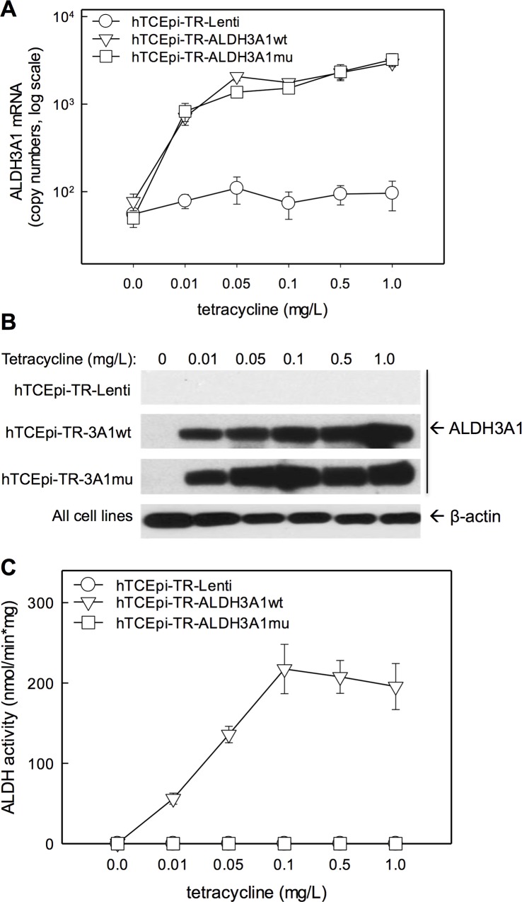
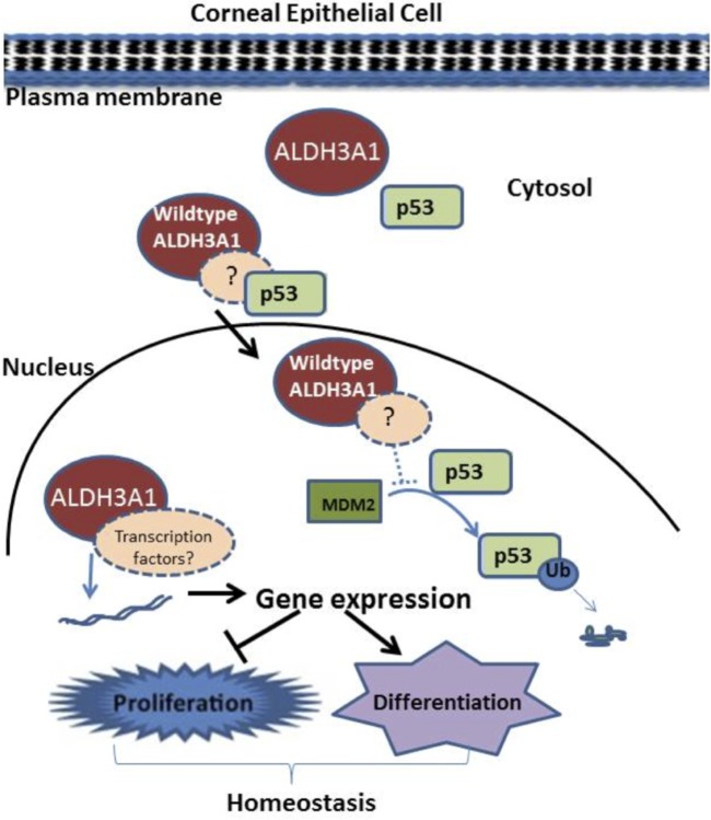
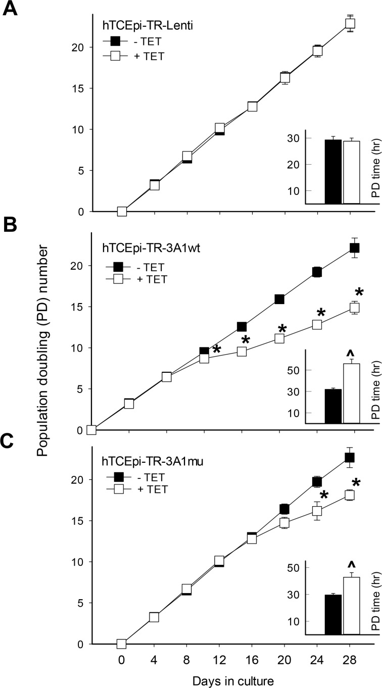
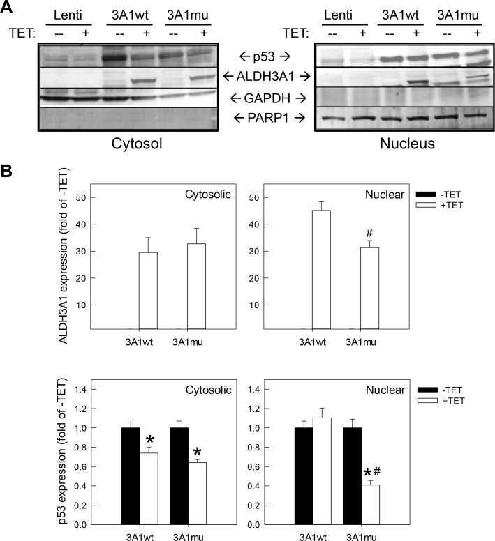
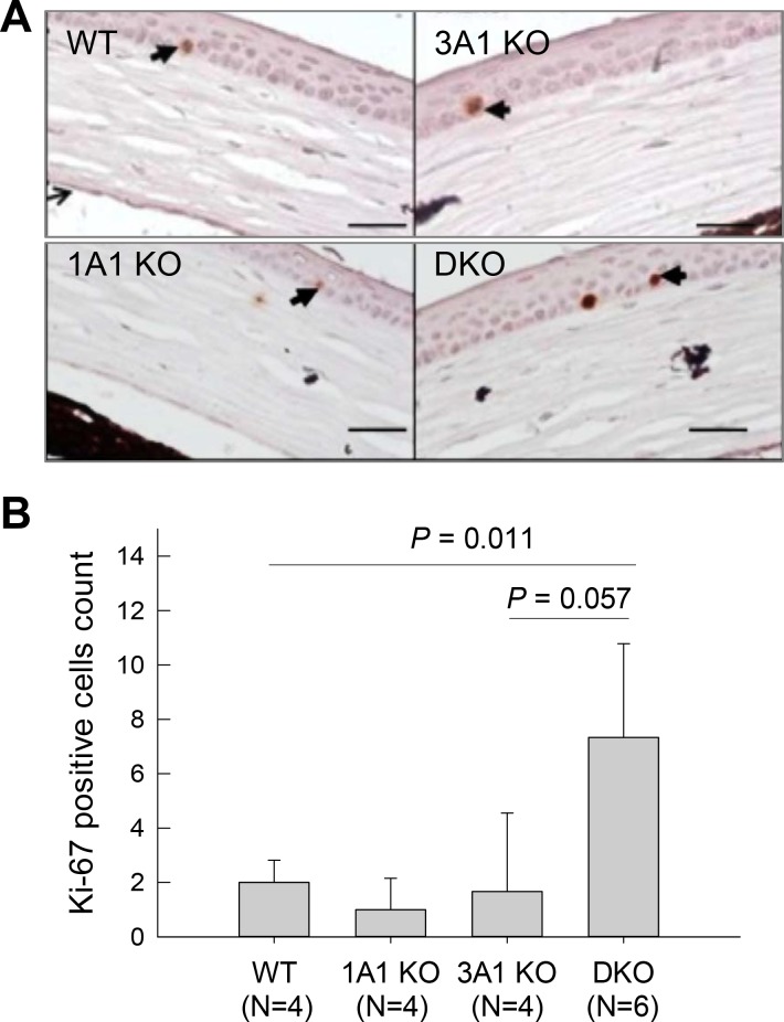
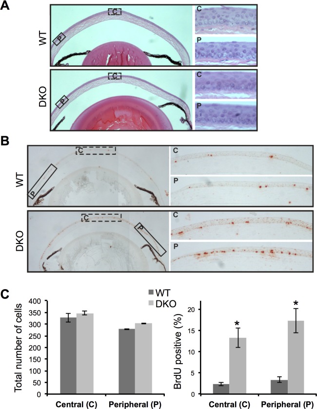
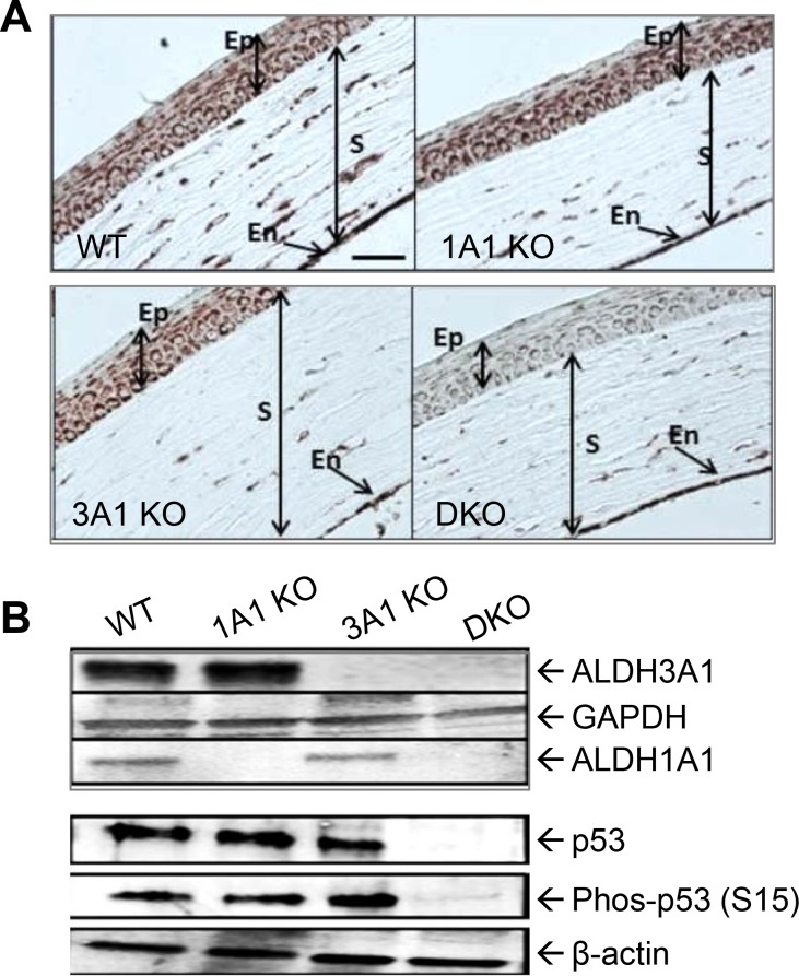
Aldehyde dehydrogenase 1A1 (ALDH1A1) and ALDH3A1 are corneal crystallins. They protect inner ocular tissues from ultraviolet radiation (UVR)-induced oxidative damage through catalytic and non-catalytic mechanisms. Additionally, ALDH3A1 has been postulated to play a regulatory role in the corneal epithelium based on several studies that report an inverse association between ALDH3A1 expression and corneal cell proliferation. The underlying molecular mechanisms and the physiological significance of such association remain poorly understood. In the current study, we established Tet-On human corneal epithelial cell (hTCEpi) lines, which express tetracycline-inducible wild-type (wt) or catalytically-inactive (mu) ALDH3A1. Utilizing this cellular model system, we confirmed that human ALDH3A1 decreases corneal cell proliferation; importantly, this effect appears to be partially mediated by its enzymatic activity. Mechanistically, wt-ALDH3A1, but not mu-ALDH3A1, promotes sequestering of tumor suppressor p53 in the nucleus. In the mouse cornea, however, augmented cell proliferation is noted only in Aldh1a1(-/-)/3a1(-/-) double knockout (DKO) mice, indicating in vivo the anti-proliferation effect of ALDH3A1 can be rescued by the presence of ALDH1A1. Interestingly, the hyper-proliferative epithelium of the DKO corneas display nearly complete loss of p53 expression, implying that p53 may be involved in ALDH3A1/1A1-mediated effect. In hTCEpi cells grown in high calcium concentration, mRNA levels of a panel of corneal differentiation markers were altered by ALDH3A1 expression and modulated by its enzyme activity. In conclusion, we show for the first time that: (i) ALDH3A1 decreases corneal epithelial proliferation through both non-enzymatic and enzymatic properties; (ii) ALDH1A1 contributes to the regulation of corneal cellular proliferation in vivo; and (iii) ALDH3A1 modulates corneal epithelial differentiation. Collectively, our studies indicate a functional role of ALDH3A1 in the maintenance of corneal epithelial homeostasis by simultaneously modulating proliferation and differentiation through both enzymatic and non-enzymatic mechanisms.
醛脱氢酶1A1(ALDH1A1)和ALDH3A1是角膜晶状体蛋白。它们通过催化和非催化机制保护眼内组织免受紫外线辐射(UVR)诱导的氧化损伤。此外,基于多项报道ALDH3A1表达与角膜细胞增殖呈负相关的研究,推测ALDH3A1在角膜上皮中起调节作用。这种关联的潜在分子机制和生理意义仍知之甚少。在本研究中,我们建立了Tet-On人角膜上皮细胞(hTCEpi)系,其表达四环素诱导型野生型(wt)或催化失活型(mu)ALDH3A1。利用这个细胞模型系统,我们证实人ALDH3A1可降低角膜细胞增殖;重要的是,这种作用似乎部分由其酶活性介导。从机制上讲,wt-ALDH3A1而非mu-ALDH3A1可促进肿瘤抑制因子p53在细胞核中的隔离。然而,在小鼠角膜中,仅在Aldh1a1(-/-)/3a1(-/-)双敲除(DKO)小鼠中观察到细胞增殖增加,这表明在体内ALDH1A1的存在可挽救ALDH3A1的抗增殖作用。有趣的是,DKO角膜的过度增殖上皮显示p53表达几乎完全丧失,这意味着p53可能参与了ALDH3A1/1A1介导的效应。在高钙浓度下生长的hTCEpi细胞中,一组角膜分化标志物的mRNA水平因ALDH3A1的表达而改变,并受其酶活性调节。总之,我们首次表明:(i)ALDH3A1通过非酶和酶特性降低角膜上皮增殖;(ii)ALDH1A1在体内有助于调节角膜细胞增殖;(iii)ALDH3A1调节角膜上皮分化。总的来说,我们的研究表明ALDH3A通过酶和非酶机制同时调节增殖和分化,在维持角膜上皮稳态中发挥功能作用。