Desai Seema S, Tung Jason C, Zhou Vivian X, Grenert James P, Malato Yann, Rezvani Milad, Español-Suñer Regina, Willenbring Holger, Weaver Valerie M, Chang Tammy T
Department of Surgery, University of California, San Francisco, San Francisco, CA.
Center for Bioengineering and Tissue Regeneration, University of California, San Francisco, San Francisco, CA.
Hepatology. 2016 Jul;64(1):261-75. doi: 10.1002/hep.28450. Epub 2016 Mar 9.
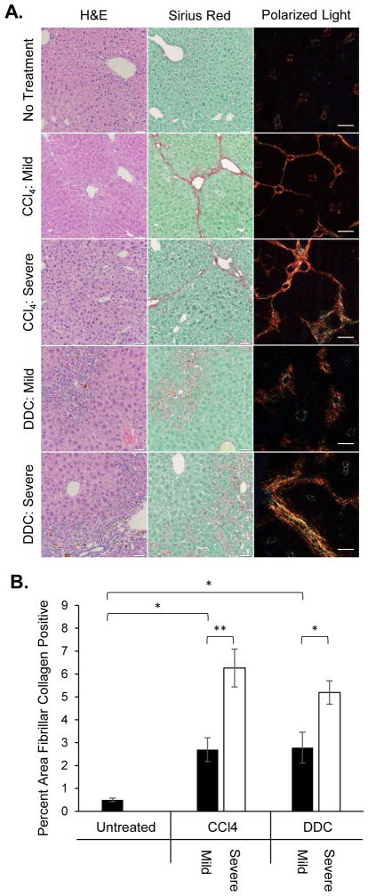
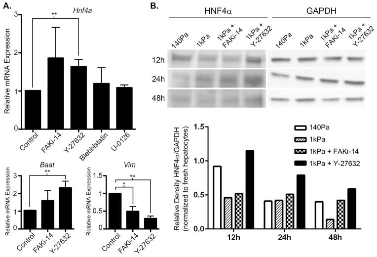
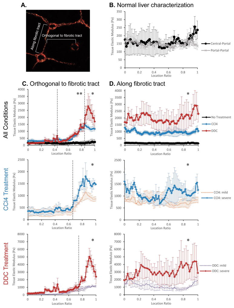
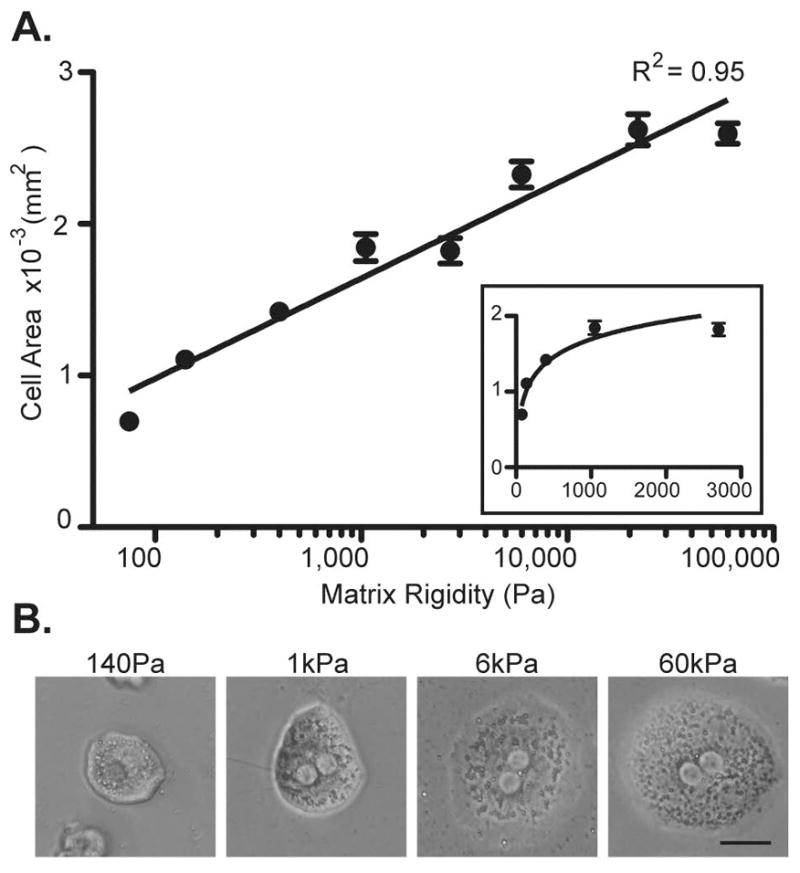
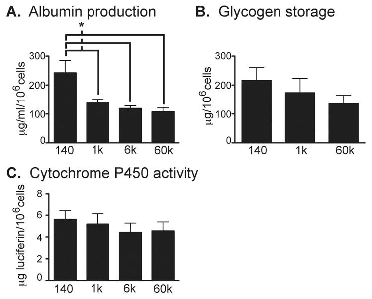
Matrix rigidity has important effects on cell behavior and is increased during liver fibrosis; however, its effect on primary hepatocyte function is unknown. We hypothesized that increased matrix rigidity in fibrotic livers would activate mechanotransduction in hepatocytes and lead to inhibition of liver-specific functions. To determine the physiologically relevant ranges of matrix stiffness at the cellular level, we performed detailed atomic force microscopy analysis across liver lobules from normal and fibrotic livers. We determined that normal liver matrix stiffness was around 150 Pa and increased to 1-6 kPa in areas near fibrillar collagen deposition in fibrotic livers. In vitro culture of primary hepatocytes on collagen matrix of tunable rigidity demonstrated that fibrotic levels of matrix stiffness had profound effects on cytoskeletal tension and significantly inhibited hepatocyte-specific functions. Normal liver stiffness maintained functional gene regulation by hepatocyte nuclear factor 4 alpha (HNF4α), whereas fibrotic matrix stiffness inhibited the HNF4α transcriptional network. Fibrotic levels of matrix stiffness activated mechanotransduction in primary hepatocytes through focal adhesion kinase. In addition, blockade of the Rho/Rho-associated protein kinase pathway rescued HNF4α expression from hepatocytes cultured on stiff matrix.
Fibrotic levels of matrix stiffness significantly inhibit hepatocyte-specific functions in part by inhibiting the HNF4α transcriptional network mediated through the Rho/Rho-associated protein kinase pathway. Increased appreciation of the role of matrix rigidity in modulating hepatocyte function will advance our understanding of the mechanisms of hepatocyte dysfunction in liver cirrhosis and spur development of novel treatments for chronic liver disease. (Hepatology 2016;64:261-275).
基质硬度对细胞行为有重要影响,且在肝纤维化过程中会增加;然而,其对原代肝细胞功能的影响尚不清楚。我们推测,纤维化肝脏中增加的基质硬度会激活肝细胞中的机械转导并导致肝脏特异性功能受到抑制。为了确定细胞水平上与生理相关的基质硬度范围,我们对正常和纤维化肝脏的肝小叶进行了详细的原子力显微镜分析。我们确定正常肝脏基质硬度约为150帕斯卡,在纤维化肝脏中纤维状胶原沉积附近区域增加到1 - 6千帕斯卡。在可调硬度的胶原基质上对原代肝细胞进行体外培养表明,纤维化水平的基质硬度对细胞骨架张力有深远影响,并显著抑制肝细胞特异性功能。正常肝脏硬度通过肝细胞核因子4α(HNF4α)维持功能基因调控,而纤维化基质硬度抑制HNF4α转录网络。纤维化水平的基质硬度通过粘着斑激酶激活原代肝细胞中的机械转导。此外,阻断Rho/ Rho相关蛋白激酶途径可挽救在硬基质上培养的肝细胞中的HNF4α表达。
纤维化水平的基质硬度通过抑制由Rho/ Rho相关蛋白激酶途径介导的HNF4α转录网络,显著抑制肝细胞特异性功能。对基质硬度在调节肝细胞功能中作用的进一步认识将推进我们对肝硬化中肝细胞功能障碍机制的理解,并推动慢性肝病新疗法的开发。(《肝脏病学》2016年;64卷:261 - 275页)