Yang Sheau-Fang, Hou Ming-Feng, Chen Fang-Ming, Ou-Yang Fu, Wu Yang-Chang, Chai Chee-Yin, Yeh Yao-Tsung
Department of Pathology, Kaohsiung Municipal Ta-Tung Hospital, No. 68, Zhonghua 3rd Rd, Qianjin Dist, Kaohsiung, 801, R O C, Taiwan.
Department of Pathology, Faculty of Medicine, College of Medicine, Kaohsiung Medical University, No.100, Shiquan 1st Rd, Sanmin Dist, Kaohsiung, 807, R O C, Taiwan.
BMC Cancer. 2016 Jan 14;16:20. doi: 10.1186/s12885-016-2063-1.
Deregulated signal transducer and activator of transcription 3 (STAT3) signaling has been well documented in certain cancers. Alterations in specific negative regulators, such as protein inhibitor of activated STAT3 (PIAS3), may contribute to cancer development.
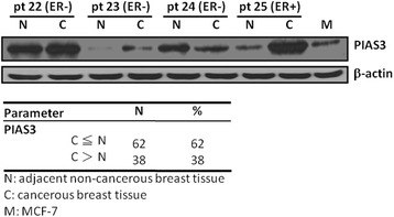
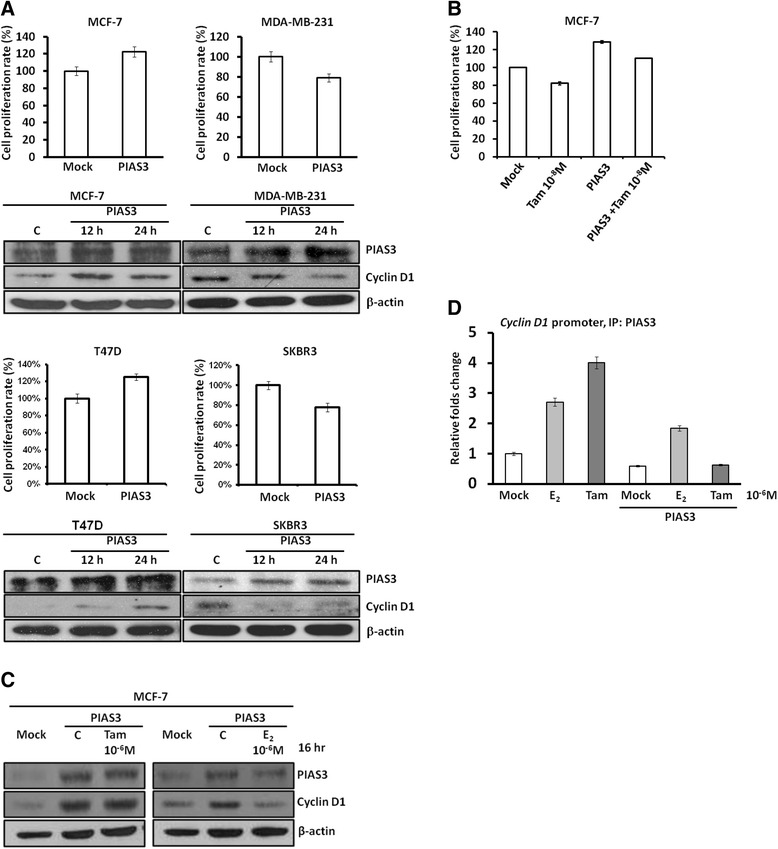
The expression of total PIAS3 was determined in 100 paired cancerous and non-cancerous breast tissues by immunoblotting and was statistically analyzed along with the clinicopathological characteristics and overall survival of the patients. XTT, immunoblotting, and chromatin immunoprecipitation (Chip) were used to examine the biological effect of PIAS3 in breast cancer cells.
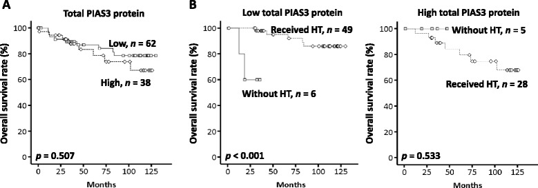
Hormone therapy failed to improve the overall survival in patients presenting with increased PIAS3 expression. Ectopic PIAS3 overexpression increased the proliferation and expression of cyclin D1 in estrogen receptor (ER)-positive MCF-7 and T47D cells, but decreased those in ER-negative MDA-MB-231 and SKBR3 cells. Furthermore, PIAS3 overexpression attenuated cytotoxicity of tamoxifen and increased proliferation and cyclin D1 expression in MCF-7 cells. PIAS3 also decreased the binding of itself on the cyclin D1 promoter and this decreased binding was not affected by tamoxifen.
PIAS3 may serve as a biomarker for predicting hormone therapy stratification, although it is limited to those breast cancer patients receiving hormone therapy.
信号转导及转录激活因子3(STAT3)信号通路失调在某些癌症中已有充分记载。特定负调控因子的改变,如活化STAT3蛋白抑制剂(PIAS3),可能有助于癌症的发展。
采用免疫印迹法检测100对乳腺癌组织和癌旁非癌组织中总PIAS3的表达,并结合患者的临床病理特征和总生存期进行统计学分析。采用XTT法、免疫印迹法和染色质免疫沉淀法(Chip)检测PIAS3在乳腺癌细胞中的生物学效应。
激素治疗未能改善PIAS3表达增加患者的总生存期。异位PIAS3过表达增加了雌激素受体(ER)阳性的MCF-7和T47D细胞的增殖及细胞周期蛋白D1的表达,但降低了ER阴性的MDA-MB-231和SKBR3细胞中的上述指标。此外,PIAS3过表达减弱了他莫昔芬的细胞毒性,并增加了MCF-7细胞的增殖和细胞周期蛋白D1的表达。PIAS3还降低了其自身与细胞周期蛋白D1启动子的结合,且这种结合的降低不受他莫昔芬的影响。
PIAS3可能作为预测激素治疗分层的生物标志物,尽管它仅限于接受激素治疗的乳腺癌患者。