Loi Florence, Córdova Luis A, Zhang Ruth, Pajarinen Jukka, Lin Tzu-hua, Goodman Stuart B, Yao Zhenyu
Department of Orthopaedic Surgery, Stanford University School of Medicine, 300 Pasteur Drive, Edwards Building, Room R116, Stanford, CA, 94305, USA.
Department of Oral and Maxillofacial Surgery, Faculty of Dentistry, University of Chile, Sergio Livingstone Polhammer 943, Independencia, 8380000, Santiago, Chile.
Stem Cell Res Ther. 2016 Jan 22;7:15. doi: 10.1186/s13287-016-0276-5.
Bone formation and remodeling are influenced by the inflammatory state of the local microenvironment. In this regard, macrophages are postulated to play a crucial role in modulating osteogenesis. However, the differential effects of macrophage subsets and their plasticity on bone formation are currently unknown.
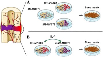
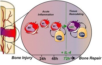
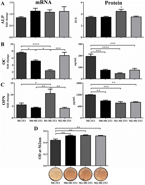
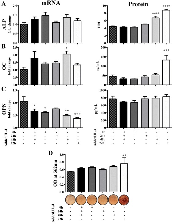
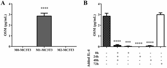
Polarized primary murine macrophages and preosteoblastic MC3T3 cells were co-cultured to investigate the effect of non-activated M0, pro-inflammatory M1, and tissue-regenerative M2 macrophages on the osteogenic ability of MC3T3-E1 cells in vitro. Furthermore, to model the physiological transition from inflammation to tissue regeneration, M1-MC3T3 co-cultures were treated with interleukin-4 (IL-4) at different time points to modulate the M1 phenotype towards M2. Macrophage phenotypic markers were assessed by flow cytometry and enzyme-linked immunosorbent assay. A time course study of osteogenic markers at different time points was conducted: alkaline phosphatase (ALP) mRNA levels were evaluated at week 1, ALP activity and osteocalcin and osteopontin mRNA levels at week 2, and matrix mineralization and osteocalcin and osteopontin protein concentrations at week 3. Supernatant collected 72 hours after seeding or IL-4 treatment, whichever was later, was analyzed for oncostatin M, a cytokine released by macrophages that has been recognized to enhance osteogenesis. Unpaired t test or one-way ANOVA with Tukey's or Dunnett's post hoc tests were used for statistical comparison of the groups.
Co-culture with any of the macrophage subtypes increased the osteogenic ability of MC3T3 cells as indicated by increases in ALP activity and matrix mineralization. Increased ALP activity, osteocalcin concentration, and matrix mineralization demonstrated that osteogenesis by M1-MC3T3 co-cultures was further enhanced by macrophage phenotype modulation to M2 via IL-4 treatment 72 hours after seeding. Increased oncostatin M protein concentration in untreated M1-MC3T3 co-cultures and M1-MC3T3 co-cultures treated with IL-4 at 72 hours correlated with greater ALP activity and matrix mineralization.
These results suggest that a transient inflammatory phase is crucial for enhanced bone formation. Macrophage plasticity may offer new strategies for modulating the local inflammatory microenvironment with the aim of potentially enhancing bone repair.
骨形成和重塑受局部微环境炎症状态的影响。在这方面,推测巨噬细胞在调节骨生成中起关键作用。然而,巨噬细胞亚群及其可塑性对骨形成的不同影响目前尚不清楚。
将极化的原代小鼠巨噬细胞与前成骨细胞MC3T3细胞共培养,以研究未活化的M0、促炎性M1和组织再生性M2巨噬细胞对MC3T3-E1细胞体外成骨能力的影响。此外,为模拟从炎症到组织再生的生理转变,在不同时间点用白细胞介素-4(IL-4)处理M1-MC3T3共培养物,以将M1表型调节为M2。通过流式细胞术和酶联免疫吸附测定评估巨噬细胞表型标志物。对不同时间点的成骨标志物进行了时间进程研究:在第1周评估碱性磷酸酶(ALP)mRNA水平,在第2周评估ALP活性、骨钙素和骨桥蛋白mRNA水平,在第3周评估基质矿化、骨钙素和骨桥蛋白蛋白浓度。在接种或IL-4处理(以较晚者为准)72小时后收集的上清液中分析制瘤素M,制瘤素M是一种由巨噬细胞释放的细胞因子,已被认为可增强骨生成。使用未配对t检验或带有Tukey或Dunnett事后检验的单向方差分析对各组进行统计学比较。
与任何巨噬细胞亚型共培养均增加了MC3T3细胞的成骨能力,表现为ALP活性和基质矿化增加。ALP活性、骨钙素浓度和基质矿化增加表明,接种72小时后通过IL-4处理将巨噬细胞表型调节为M2可进一步增强M1-MC3T3共培养物的骨生成。未处理的M1-MC3T3共培养物和在72小时用IL-4处理的M1-MC3T3共培养物中制瘤素M蛋白浓度增加与更高的ALP活性和基质矿化相关。
这些结果表明,短暂的炎症期对增强骨形成至关重要。巨噬细胞可塑性可能为调节局部炎症微环境提供新策略,以期潜在地增强骨修复。