Yang Lian, Xiao Ming, Li Xian, Tang Yi, Wang Ya-Lan
Department of Pathology, Molecular Medicine and Cancer Research Center, Chongqing Medical University, Chongqing 400016, P.R. China.
Int J Mol Med. 2016 Mar;37(3):734-42. doi: 10.3892/ijmm.2016.2473. Epub 2016 Jan 29.
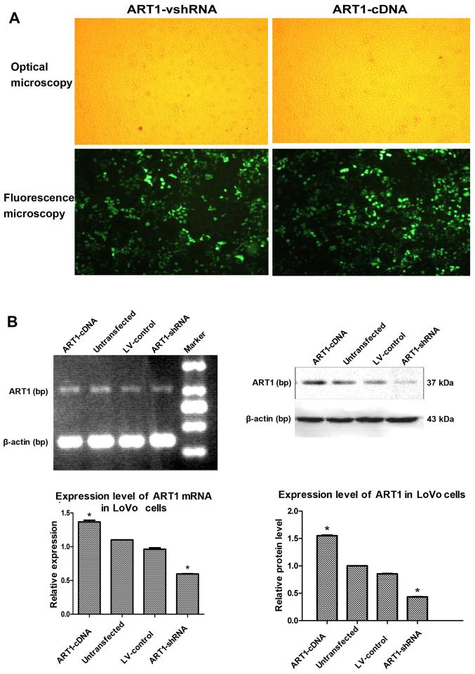
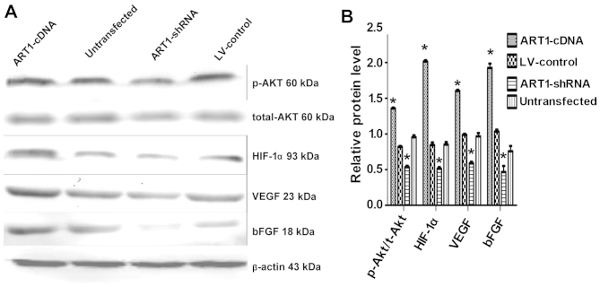
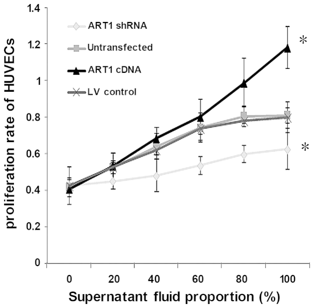
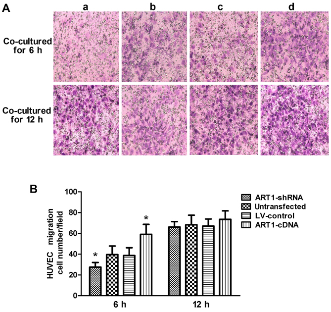
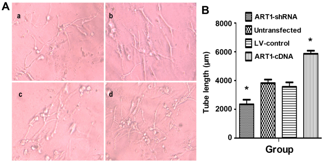
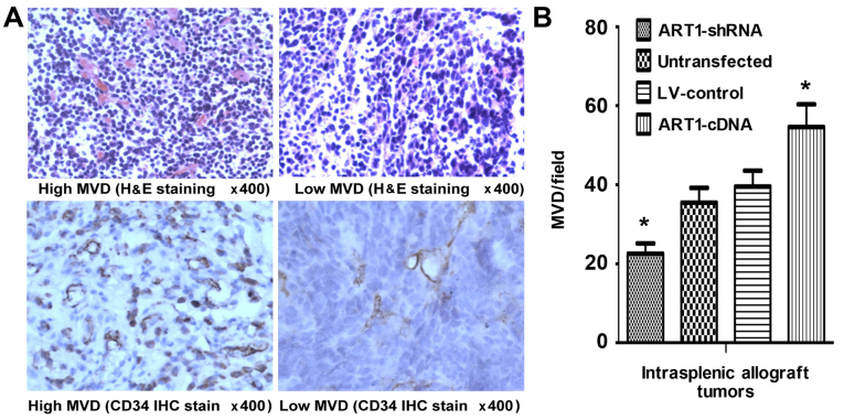
Arginine adenosine diphosphate (ADP)-ribosyl-transferase 1 (ART1) is known to play an important role in many physiological and pathological processes. Previous studies have demonstrated that ART1 promotes proliferation, invasion and metastasis in colon carcinoma. However, it was unclear whether ART1 is involved in angiogenesis in cases of colorectal cancer (CRC). In the present study, lentiviral vector‑mediated ART1‑cDNA or ART1-shRNA were transfected into LoVo cells, and the LoVo cells transfected with ART1-cDNA or ART1-shRNA were co-cultured with human umbilical vein endothelial cells (HUVECs) to determine the influence of ART1 on HUVECs. The proliferation, migration and angiogenesis of HUVECs were monitored using a cell counting kit-8 assay, a Transwell migration assay and immunohistochemical analysis in intrasplenic allograft tumors, respectively. Hypoxia‑inducible factor 1-α (HIF-1α), total (t-)Akt, phosphorylated (p-)Akt, vascular endothelial growth factor (VEGF) and basic fibroblast growth factor (bFGF) expression levels were detected via western blot analysis. Our results revealed that HUVECs which were co-cultured with ART1-cDNA LoVo cells showed higher proliferation, migration and angiogenic abilities, but a reduction was noted in those cultured with ART1-shRNA LoVo cells; p-Akt, HIF-1α, VEGF and bFGF expression was increased in HUVECs cultured with ART1‑cDNA-transfected LoVo cells, but reduced in ART1-shRNA-transfected LoVo cells. In a mouse xenograft model, we noted that the tumor microvessel density (MVD) was significantly increased in intrasplenic transplanted ART1‑cDNA CT26 tumors but decreased in intrasplenic transplanted ART1‑shRNA tumors. These data suggest that ART1 promoted the expression of HIF-1α via the Akt pathway in tumor cells. It also upregulated VEGF and bFGF and enhanced angiogenesis in HUVECs. Thus, we suggest that ART1 plays an important role in the invasion of CRC cells and the metastasis of CRC.
已知精氨酸二磷酸(ADP)核糖基转移酶1(ART1)在许多生理和病理过程中发挥重要作用。先前的研究表明,ART1促进结肠癌的增殖、侵袭和转移。然而,尚不清楚ART1是否参与结直肠癌(CRC)的血管生成。在本研究中,将慢病毒载体介导的ART1-cDNA或ART1-shRNA转染到LoVo细胞中,并将转染了ART1-cDNA或ART1-shRNA的LoVo细胞与人脐静脉内皮细胞(HUVECs)共培养,以确定ART1对HUVECs的影响。分别使用细胞计数试剂盒-8检测、Transwell迁移检测和脾内移植瘤的免疫组织化学分析来监测HUVECs的增殖、迁移和血管生成。通过蛋白质印迹分析检测缺氧诱导因子1-α(HIF-1α)、总(t-)Akt、磷酸化(p-)Akt、血管内皮生长因子(VEGF)和碱性成纤维细胞生长因子(bFGF)的表达水平。我们的结果显示,与ART1-cDNA LoVo细胞共培养的HUVECs表现出更高的增殖、迁移和血管生成能力,但与ART1-shRNA LoVo细胞共培养的HUVECs则出现降低;与转染ART1-cDNA的LoVo细胞共培养的HUVECs中,p-Akt、HIF-1α、VEGF和bFGF表达增加,但在转染ART1-shRNA的LoVo细胞中降低。在小鼠异种移植模型中,我们注意到脾内移植ART1-cDNA CT26肿瘤的肿瘤微血管密度(MVD)显著增加,但脾内移植ART1-shRNA肿瘤的MVD降低。这些数据表明,ART1通过肿瘤细胞中的Akt途径促进HIF-1α的表达。它还上调VEGF和bFGF,并增强HUVECs中的血管生成。因此,我们认为ART1在CRC细胞的侵袭和CRC的转移中起重要作用。