Macey Paul M, Ogren Jennifer A, Kumar Rajesh, Harper Ronald M
UCLA School of Nursing, University of California at Los AngelesLos Angeles, CA, USA; Brain Research Institute, University of California at Los AngelesLos Angeles, CA, USA.
Department of Neurobiology, University of California at Los Angeles Los Angeles, CA, USA.
Front Neurosci. 2016 Jan 26;9:513. doi: 10.3389/fnins.2015.00513. eCollection 2015.
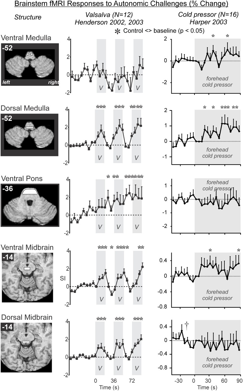
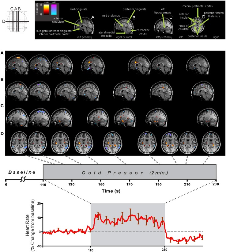
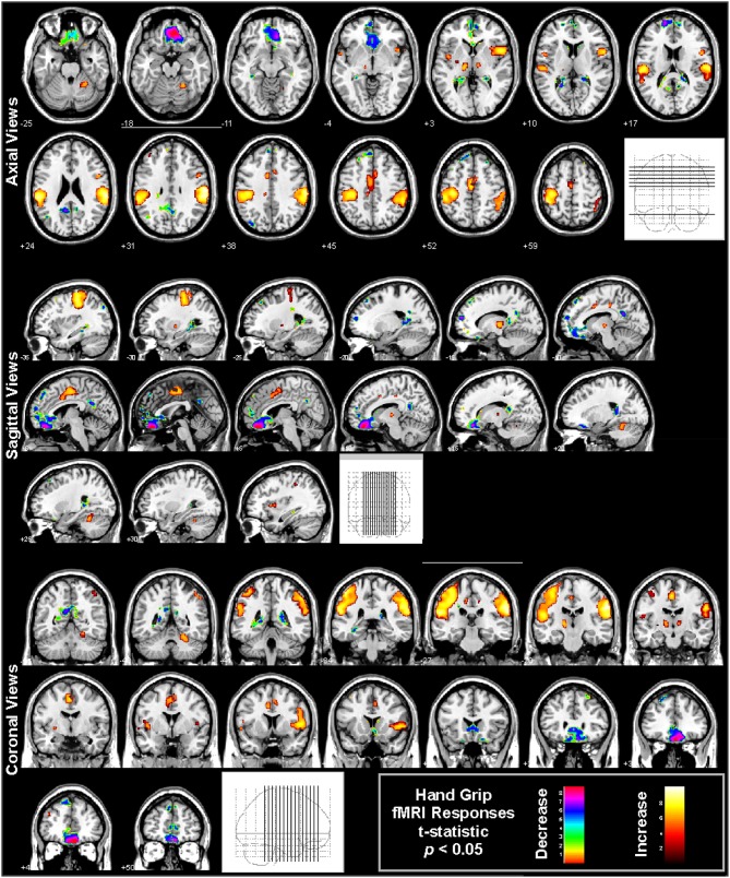
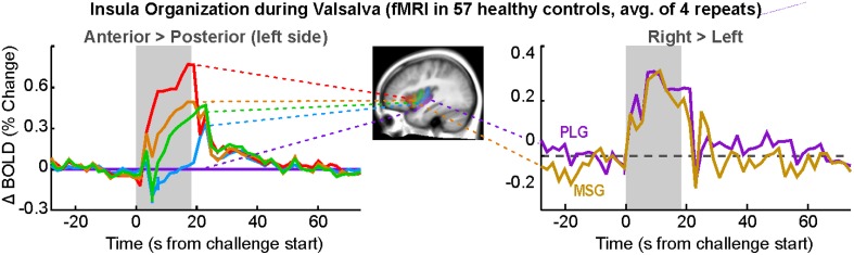
Central nervous system processing of autonomic function involves a network of regions throughout the brain which can be visualized and measured with neuroimaging techniques, notably functional magnetic resonance imaging (fMRI). The development of fMRI procedures has both confirmed and extended earlier findings from animal models, and human stroke and lesion studies. Assessments with fMRI can elucidate interactions between different central sites in regulating normal autonomic patterning, and demonstrate how disturbed systems can interact to produce aberrant regulation during autonomic challenges. Understanding autonomic dysfunction in various illnesses reveals mechanisms that potentially lead to interventions in the impairments. The objectives here are to: (1) describe the fMRI neuroimaging methodology for assessment of autonomic neural control, (2) outline the widespread, lateralized distribution of function in autonomic sites in the normal brain which includes structures from the neocortex through the medulla and cerebellum, (3) illustrate the importance of the time course of neural changes when coordinating responses, and how those patterns are impacted in conditions of sleep-disordered breathing, and (4) highlight opportunities for future research studies with emerging methodologies. Methodological considerations specific to autonomic testing include timing of challenges relative to the underlying fMRI signal, spatial resolution sufficient to identify autonomic brainstem nuclei, blood pressure, and blood oxygenation influences on the fMRI signal, and the sustained timing, often measured in minutes of challenge periods and recovery. Key findings include the lateralized nature of autonomic organization, which is reminiscent of asymmetric motor, sensory, and language pathways. Testing brain function during autonomic challenges demonstrate closely-integrated timing of responses in connected brain areas during autonomic challenges, and the involvement with brain regions mediating postural and motoric actions, including respiration, and cardiac output. The study of pathological processes associated with autonomic disruption shows susceptibilities of different brain structures to altered timing of neural function, notably in sleep disordered breathing, such as obstructive sleep apnea and congenital central hypoventilation syndrome. The cerebellum, in particular, serves coordination roles for vestibular stimuli and blood pressure changes, and shows both injury and substantially altered timing of responses to pressor challenges in sleep-disordered breathing conditions. The insights into central autonomic processing provided by neuroimaging have assisted understanding of such regulation, and may lead to new treatment options for conditions with disrupted autonomic function.
自主神经系统功能的中枢神经系统处理涉及大脑中多个区域组成的网络,这些区域可以通过神经成像技术,尤其是功能磁共振成像(fMRI)进行可视化和测量。fMRI程序的发展既证实并扩展了早期动物模型、人类中风及病变研究的结果。fMRI评估能够阐明不同中枢位点在调节正常自主神经模式时的相互作用,并展示在自主神经应激期间,紊乱的系统如何相互作用以产生异常调节。了解各种疾病中的自主神经功能障碍揭示了可能导致对这些损伤进行干预的机制。这里的目标是:(1)描述用于评估自主神经控制的fMRI神经成像方法;(2)概述正常大脑中自主神经位点广泛的、呈侧化分布的功能,这些位点包括从新皮质到延髓和小脑的结构;(3)说明神经变化的时间进程在协调反应时的重要性,以及这些模式在睡眠呼吸障碍情况下如何受到影响;(4)强调利用新兴方法进行未来研究的机会。自主神经测试特有的方法学考虑因素包括相对于潜在fMRI信号的应激时间、足以识别自主神经脑干核的空间分辨率、血压和血液氧合对fMRI信号的影响,以及通常以应激期和恢复期的分钟数来衡量的持续时间。主要发现包括自主神经组织的侧化性质,这让人联想到不对称的运动、感觉和语言通路。在自主神经应激期间测试脑功能表明,在自主神经应激期间,相连脑区的反应时间紧密整合,并且涉及介导姿势和运动动作(包括呼吸和心输出量)的脑区。对与自主神经破坏相关的病理过程的研究表明,不同脑结构对神经功能时间改变的易感性,尤其是在睡眠呼吸障碍中,如阻塞性睡眠呼吸暂停和先天性中枢性低通气综合征。特别是小脑,它在前庭刺激和血压变化中起协调作用,并且在睡眠呼吸障碍情况下,对升压应激的反应显示出损伤和反应时间的显著改变。神经成像对中枢自主神经处理的深入了解有助于理解这种调节,并可能为自主神经功能紊乱的疾病带来新的治疗选择。