Haas Stefan Jean-Pierre, Zhou Xiaolai, Machado Venissa, Wree Andreas, Krieglstein Kerstin, Spittau Björn
Institute of Anatomy, Rostock University Medical Center Rostock, Germany.
Department of Molecular Embryology, Institute of Anatomy and Cell Biology, Albert-Ludwigs-UniversityFreiburg, Germany; Department of Molecular Biology and Genetics, Weill Institute for Cell and Molecular Biology, Cornell UniversityIthaca, NY, USA.
Front Mol Neurosci. 2016 Feb 3;9:7. doi: 10.3389/fnmol.2016.00007. eCollection 2016.
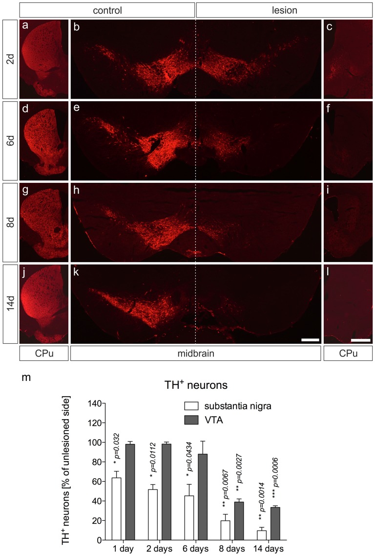
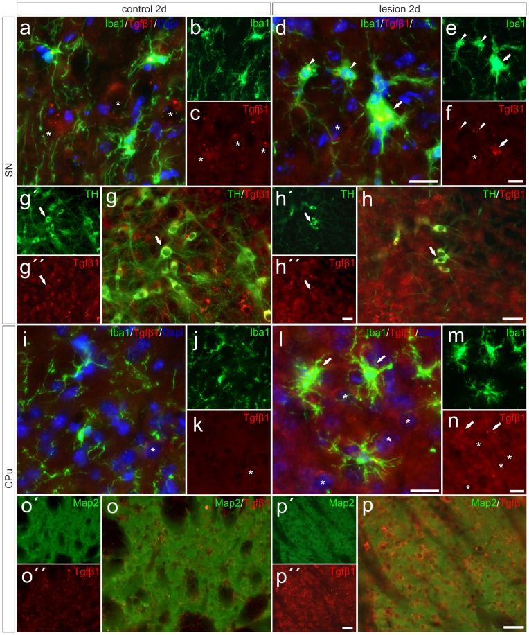
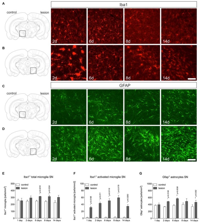
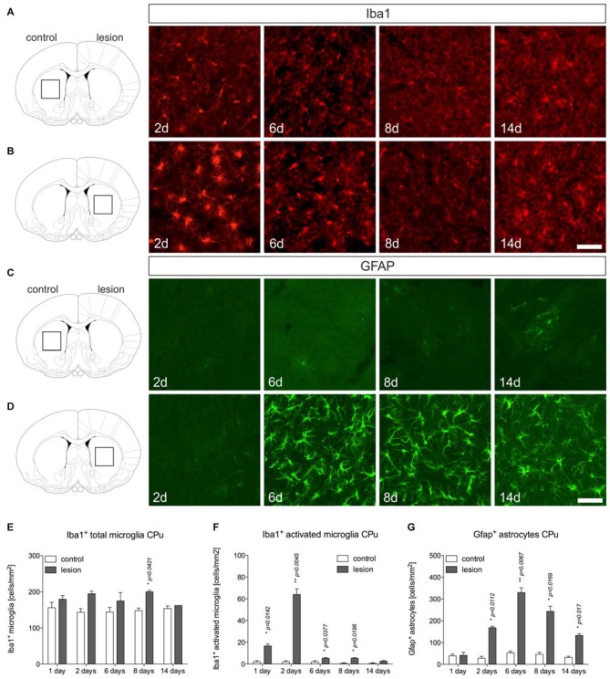
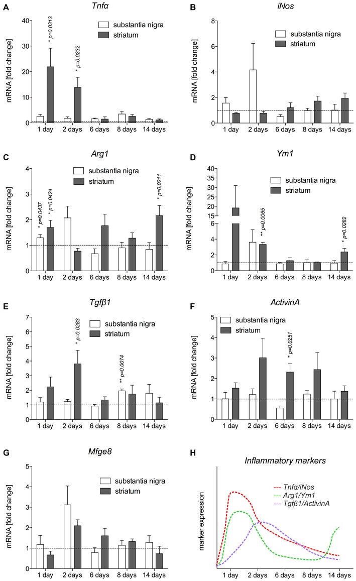
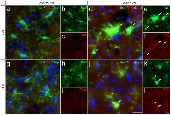
Parkinson's disease (PD) is a neurodegenerative disorder that is characterized by loss of midbrain dopaminergic (mDA) neurons in the substantia nigra (SN). Microglia-mediated neuroinflammation has been described as a common hallmark of PD and is believed to further trigger the progression of neurodegenerative events. Injections of 6-hydroxydopamine (6-OHDA) are widely used to induce degeneration of mDA neurons in rodents as an attempt to mimic PD and to study neurodegeneration, neuroinflammation as well as potential therapeutic approaches. In the present study, we addressed microglia and astroglia reactivity in the SN and the caudatoputamen (CPu) after 6-OHDA injections into the medial forebrain bundle (MFB), and further analyzed the temporal and spatial expression patterns of pro-inflammatory and anti-inflammatory markers in this mouse model of PD. We provide evidence that activated microglia as well as neurons in the lesioned SN and CPu express Transforming growth factor β1 (Tgfβ1), which overlaps with the downregulation of pro-inflammatory markers Tnfα, and iNos, and upregulation of anti-inflammatory markers Ym1 and Arg1. Taken together, the data presented in this study suggest an important role for Tgfβ1 as a lesion-associated factor that might be involved in regulating microglia activation states in the 6-OHDA mouse model of PD in order to prevent degeneration of uninjured neurons by microglia-mediated release of neurotoxic factors such as Tnfα and nitric oxide (NO).
帕金森病(PD)是一种神经退行性疾病,其特征是黑质(SN)中脑多巴胺能(mDA)神经元丧失。小胶质细胞介导的神经炎症被认为是PD的一个常见特征,并且被认为会进一步引发神经退行性事件的进展。注射6-羟基多巴胺(6-OHDA)被广泛用于诱导啮齿动物中mDA神经元变性,以试图模拟PD并研究神经退行性变、神经炎症以及潜在的治疗方法。在本研究中,我们研究了向内侧前脑束(MFB)注射6-OHDA后,SN和尾状壳核(CPu)中小胶质细胞和星形胶质细胞的反应性,并进一步分析了该PD小鼠模型中促炎和抗炎标志物的时空表达模式。我们提供的证据表明,在受损的SN和CPu中,活化的小胶质细胞以及神经元表达转化生长因子β1(Tgfβ1),这与促炎标志物Tnfα和诱导型一氧化氮合酶(iNos)的下调以及抗炎标志物Ym1和精氨酸酶1(Arg1)的上调重叠。综上所述,本研究中的数据表明Tgfβ1作为一种损伤相关因子具有重要作用,它可能参与调节6-OHDA诱导的PD小鼠模型中的小胶质细胞活化状态,以防止未受损神经元因小胶质细胞介导释放神经毒性因子如Tnfα和一氧化氮(NO)而发生变性。