Iacobellis Francesca, Brillantino Antonio, Renzi Adolfo, Monaco Luigi, Serra Nicola, Feragalli Beatrice, Iacomino Aniello, Brunese Luca, Cappabianca Salvatore
Department of Radiology, Second University of Naples, Piazza Miraglia 2, 80138 Napoli, Italy.
"Villa delle Querce" Hospital, Via Battistello Caracciolo 48, 80136 Napoli, Italy.
Gastroenterol Res Pract. 2016;2016:6594152. doi: 10.1155/2016/6594152. Epub 2016 Jan 12.
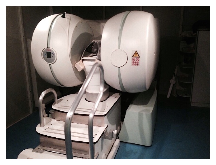
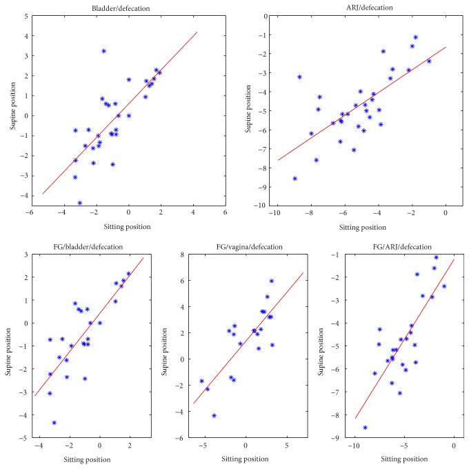
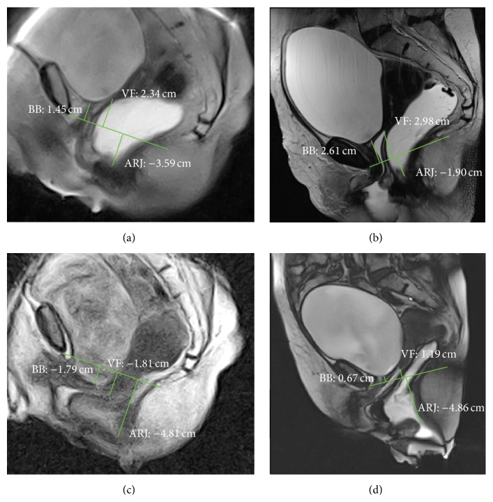
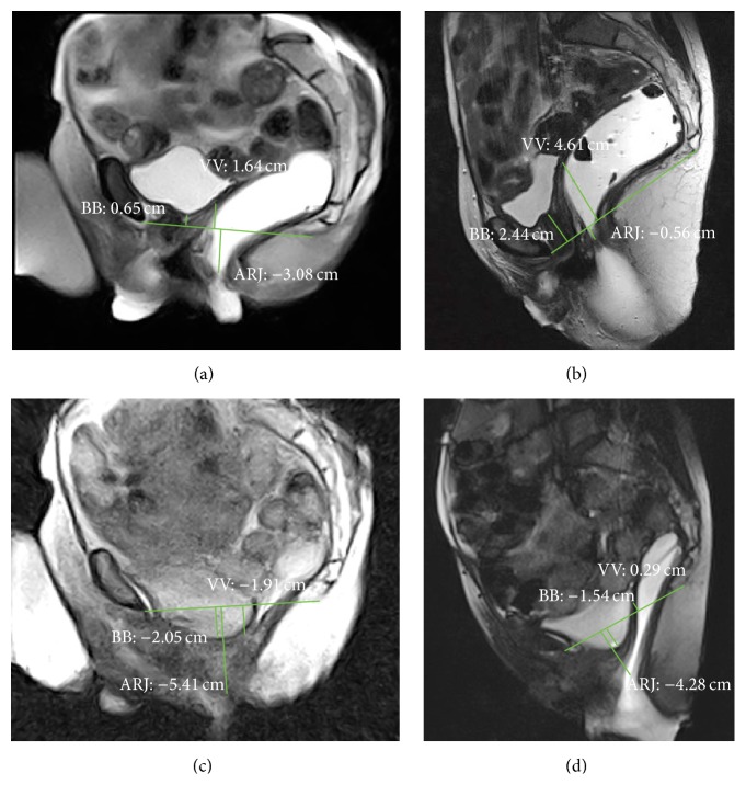
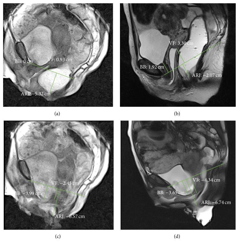
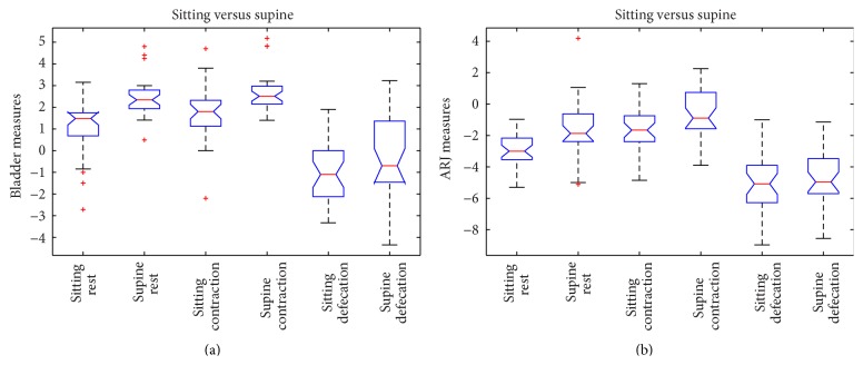
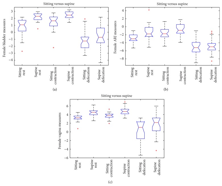
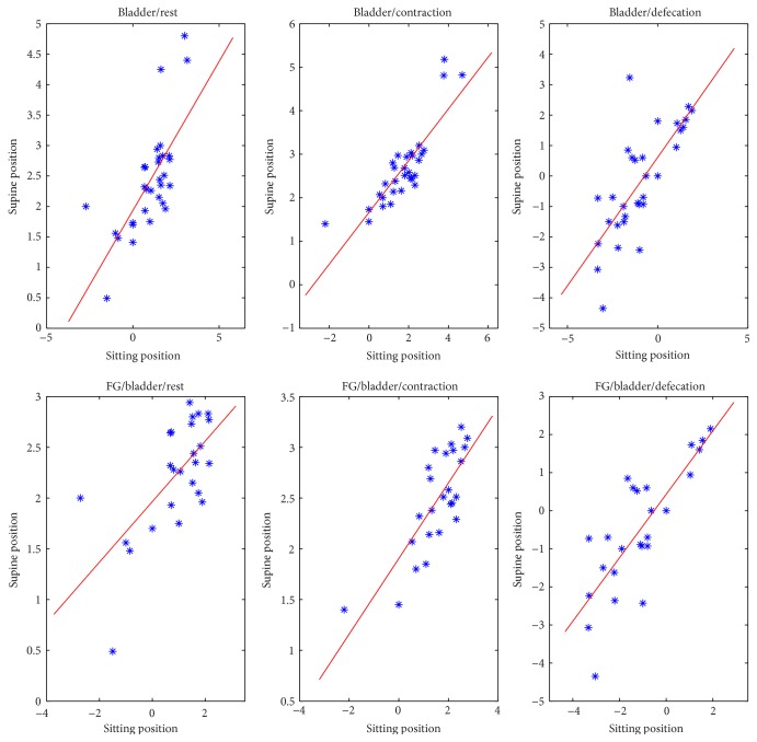
Introduction. Functional disorders of the pelvic floor represent have a significant impact on the quality of life. The advent of open-configuration systems allowed for the evaluation of defecation with MR imaging in sitting position. The purpose of the present study is to compare the results of static and dynamic pelvic MR performed in supine position versus sitting position, using a new MR prototype machine, in the diagnosis of pelvic floor descent. Materials and Methods. Thirty-one patients with pelvic floor disorders were enrolled, and underwent MR Defecography in supine position with 1.5 T closed magnet (MAGNETOM Symphony, Siemens, Germany) and in sitting position with a 0.25-Tesla open magnet system (G-Scan ESAOTE, Italy). Results. In rest and squeezing phases, positions of bladder, vagina, and ARJ were significantly different when the patient was imaged in supine versus sitting position. In the defecation phase, a significant difference for the bladder and vagina position was detected between the two exams whereas a significant difference for the ARJ was not found. A statistically significant difference exists when the pelvic floor descent is evaluated in sitting versus supine position. Conclusion. Our results show that MR Defecography in sitting position may represent a useful tool to correctly diagnose and grade the pelvic organ descent.
引言。盆底功能障碍对生活质量有重大影响。开放式系统的出现使得在坐位时利用磁共振成像评估排便情况成为可能。本研究的目的是使用新型磁共振原型机,比较仰卧位与坐位时进行的静态和动态盆腔磁共振成像在诊断盆底下降方面的结果。材料与方法。招募了31例盆底功能障碍患者,分别在1.5T封闭磁体(德国西门子MAGNETOM Symphony)下仰卧位以及0.25T开放式磁体系统(意大利ESAOTE G-Scan)下坐位接受磁共振排粪造影检查。结果。在静息期和挤压期,患者仰卧位与坐位成像时膀胱、阴道和肛管直肠连接部的位置存在显著差异。在排便期,两次检查之间膀胱和阴道位置存在显著差异,而肛管直肠连接部未发现显著差异。在坐位与仰卧位评估盆底下降时存在统计学显著差异。结论。我们的结果表明,坐位磁共振排粪造影可能是正确诊断和分级盆腔器官下降的有用工具。