Eftekhar-Vaghefi Shahrzad, Esmaeili-Mahani Saeed, Elyasi Leila, Abbasnejad Mehdi
Department of Biology, Faculty of Sciences, Shahid Bahonar University of Kerman, Kerman, Iran.; Laboratory of Molecular Neuroscience, Neuroscience Research Center, Institute of Neuropharmacology, Kerman University of Medical Sciences, Kerman, Iran.
Department of Biology, Faculty of Sciences, Shahid Bahonar University of Kerman, Kerman, Iran.
Basic Clin Neurosci. 2015 Jul;6(3):171-8.
The neuroprotective role of opioid morphine against 6-hydroxydopamine-induced cell death has been demonstrated. However, the exact mechanism(s) underlying such neuroprotection, especially the role of subtype receptors, has not yet been fully clarified.
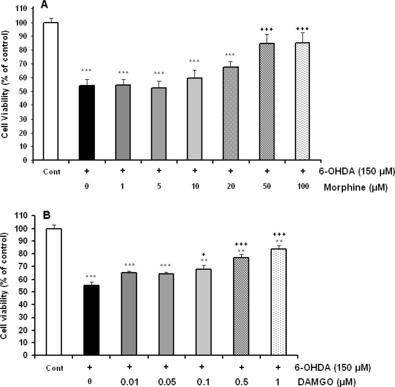
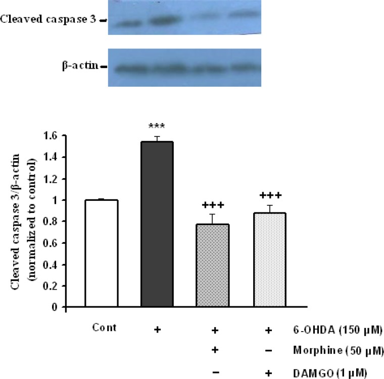
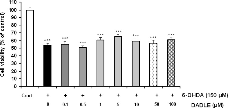
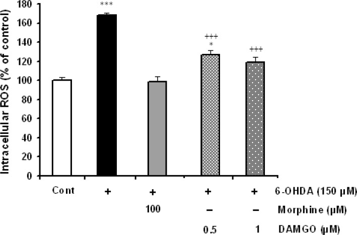
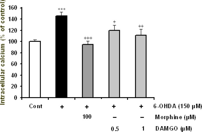
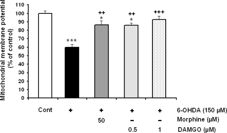
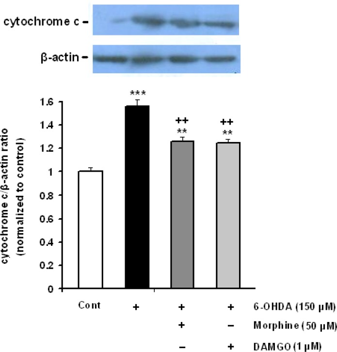
Here, we investigated the effects of different opioid agonists on 6-OHDA-induced neurotoxicity in human neuroblastoma SH-SY5Y cell line as an in vitro model of Parkinson's disease. Cell damage was induced by 150 μM 6-OHDA and the cells viability was examined by MTT assay. Intracellular calcium, reactive oxygen species and mitochondrial membrane potential were assessed by fluorescence spectrophotometry method. Immunoblot technique was used to evaluate cytochrome-c and activated caspase-3 as biochemical markers of apoptosis induction.
The data showed that 6-OHDA caused significant cell damage, loss of mitochondrial membrane potential and increase in intracellular reactive oxygen species and calcium levels as well as activated caspase-3 and cytochrome-c release. Incubation of SH-SY5Y cells with μ-opioid agonists, morphine and DAMGO, but not with δ-opioid agonist, DADLE, elicited protective effect and reduced biochemical markers of cell damage and death.
The results suggest that μ-opioid receptors signaling participate in the opioid neuroprotective effects against 6-OHDA-induced neurotoxicity.
阿片类药物吗啡对6-羟基多巴胺诱导的细胞死亡具有神经保护作用已得到证实。然而,这种神经保护作用的确切机制,尤其是亚型受体的作用,尚未完全阐明。
在此,我们研究了不同阿片类激动剂对人神经母细胞瘤SH-SY5Y细胞系中6-OHDA诱导的神经毒性的影响,该细胞系作为帕金森病的体外模型。用150μM 6-OHDA诱导细胞损伤,并通过MTT法检测细胞活力。采用荧光分光光度法评估细胞内钙、活性氧和线粒体膜电位。免疫印迹技术用于评估细胞色素c和活化的半胱天冬酶-3,作为凋亡诱导的生化标志物。
数据显示,6-OHDA导致显著的细胞损伤、线粒体膜电位丧失、细胞内活性氧和钙水平升高,以及活化的半胱天冬酶-3和细胞色素c释放。用μ-阿片类激动剂吗啡和DAMGO孵育SH-SY5Y细胞,但不用δ-阿片类激动剂DADLE孵育,可产生保护作用,并降低细胞损伤和死亡的生化标志物。
结果表明,μ-阿片类受体信号传导参与了阿片类药物对6-OHDA诱导的神经毒性的神经保护作用。